. 2011 Nov;1(3):110012.
doi: 10.1098/rsob.110012.

Discovery of catalytically active orthologues of the Parkinson's disease kinase PINK1: analysis of substrate specificity and impact of mutations

Affiliations

Discovery of catalytically active orthologues of the Parkinson's disease kinase PINK1: analysis of substrate specificity and impact of mutations

Helen I Woodroof et al. Open Biol. 2011 Nov.

Abstract

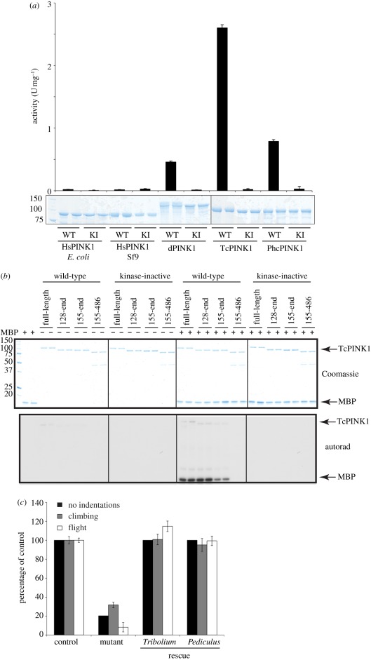
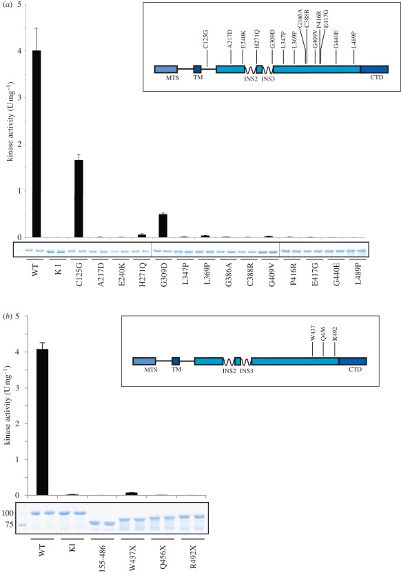
Missense mutations of the phosphatase and tensin homolog (PTEN)-induced kinase 1 (PINK1) gene cause autosomal-recessive Parkinson's disease. To date, little is known about the intrinsic catalytic properties of PINK1 since the human enzyme displays such low kinase activity in vitro. We have discovered that, in contrast to mammalian PINK1, insect orthologues of PINK1 we have investigated-namely Drosophila melanogaster (dPINK1), Tribolium castaneum (TcPINK1) and Pediculus humanus corporis (PhcPINK1)-are active as judged by their ability to phosphorylate the generic substrate myelin basic protein. We have exploited the most active orthologue, TcPINK1, to assess its substrate specificity and elaborated a peptide substrate (PINKtide, KKWIpYRRSPRRR) that can be employed to quantify PINK1 kinase activity. Analysis of PINKtide variants reveal that PINK1 phosphorylates serine or threonine, but not tyrosine, and we show that PINK1 exhibits a preference for a proline at the +1 position relative to the phosphorylation site. We have also, for the first time, been able to investigate the effect of Parkinson's disease-associated PINK1 missense mutations, and found that nearly all those located within the kinase domain, as well as the C-terminal non-catalytic region, markedly suppress kinase activity. This emphasizes the crucial importance of PINK1 kinase activity in preventing the development of Parkinson's disease. Our findings will aid future studies aimed at understanding how the activity of PINK1 is regulated and the identification of physiological substrates.

Keywords: biochemistry, Parkinson's disease, kinase.


Figures

Figure 1.
Figure 1.
Identification of orthologues of PINK1. (a) Multiple sequence alignment of PINK1 from human, T. castaneum, P. humanus corporis and D. melanogaster. The start and the end of the kinase domain are indicated by red arrows, and the three insertions within the kinase domain discussed in the text are marked with blue bars. (b) Schematic of domain structure of PINK1 orthologues in human, T. castaneum, P. humanus corporis and D. melanogaster, with numbering corresponding to the human sequence. MTS, mitochondrial targeting sequence; TM, transmembrane helix; INS, insertion; CTD, C-terminal domain.
Figure 2.
Figure 2.
Characterization of active insect orthologues of PINK1. (a) Assessment of activity of wild-type N-terminally truncated human PINK1 (125–581) expressed in E. coli and Sf9 cells, full-length D. melanogaster PINK1 (dPINK1, 1–721), T. castaneum PINK1 (TcPINK1, 1–570) and P. humanus corporis PINK1 (PhcPINK1, 1–575), and corresponding kinase-inactive mutants (HsPINK1-D384A, dPINK1-D501A, TcPINK1-D359A, PhcPINK1-D357A) against myelin basic protein (MBP). The indicated enzymes (1 µg) were incubated in the presence of 5 µg MBP and [γ-32P] ATP for 30 min. Reactions were terminated by spotting on P81 paper, washing in phosphoric acid and quantifying phosphorylation of myelin basic protein. The results are presented as ±s.d. for a representative experiment undertaken in duplicate (upper panel). In the lower panel, representative Coomassie-stained gels showing the relative amounts of PINK1 enzyme used for each assay are shown. Fine dividing lines indicate that reactions were resolved on separate gels and grouped in the final figure. (b) Assessment of kinase activity of wild-type or kinase inactive (D359A) full-length (1–570), N-terminal truncation (128–570 and 155–570) and N- and C-terminal truncation mutants (155–486) of TcPINK1. The indicated forms of TcPINK1 (1 µg) were incubated in the presence (+) or absence (−) of myelin basic protein (2 µM) and [γ-32P] ATP for 30 min. Reactions were terminated by the addition of SDS sample buffer and separated by SDS-PAGE. Gels were analysed by Coomassie staining (upper panel) and incorporation of [γ-32P] ATP was detected by autoradiography (lower panel). Fine dividing lines indicate that reactions were resolved on separate gels and grouped in the final figure. (c) Analysis of T. castaneum and P. humanus corporis PINK1 function in vivo. TcPINK1 or PhcPINK1 was ectopically expressed in Drosophila lacking endogenous PINK1. Flight ability, climbing ability and presence of thoracic indentations were quantified. Genotypes are as follows. Control: PINK1B9/+, mutant: PINK1B9/Y; da-GAL4/+, mutant rescue: PINK1B9/Y; da-GAL4/+, UAS-Tb.PINK12a/+ or PINK1B9/Y; da-GAL4/+, UAS-Phc.PINK11/+. Data are presented as mean ± s.e.m.
Figure 3.
Figure 3.
Elaboration of the PINKtide substrate for TcPINK1. (a) Full-length (1–570) (i) wild-type and (ii) kinase-inactive (D359A). TcPINK1 was used to screen a positional scanning peptide library of 198 biotinylated peptide libraries. Reaction products were bound to a streptavidin-coated membrane, washed and visualized by phospho-imaging. The small red dots indicate the residues selected for the PINKtide peptide sequence. (b) Kinetics of phosphorylation of PINKtide and indicated variants by full-length TcPINK1 (1–570). Residues that were changed relative to PINKtide are indicated in bold. Km and Vmax values were derived by nonlinear regression analysis as described in §5. n.d. denotes that a particular peptide was phosphorylated poorly and kinetic values were not determinable. Similar results were obtained in at least two experiments. (c) Kinetics of phosphorylation of mutants of PINKtide at the +1 position by full-length TcPINK1 (1–570) in a representative experiment to illustrate the marked preference for a +1 Pro residue.
Figure 4.
Figure 4.
Effect of Parkinson's disease mutation on PINK1 kinase activity. (a) Inset: Schematic of the location of missense PINK1 mutations where the wild-type residue is conserved in both human PINK1 and TcPINK1. Numbering is according to human PINK1. Mutations were introduced into full-length TcPINK1 (1–570), and enzymes (1 µg) were incubated in presence of PINKtide (1 mM) and [γ-32P] ATP for 30 min. Reactions were terminated by spotting onto P81 paper, washing in phosphoric acid and quantifying phosphorylation of PINKtide bound to P81 paper. The results are presented as ±s.d. for three experiments undertaken in duplicate. Representative Coomassie-stained gels showing the relative amounts of PINK1 enzyme used for each assay are shown. (b) Inset: Schematic of the location of C-terminally truncating PINK1 mutations. Numbering is according to human PINK1. Mutations were introduced into full-length TcPINK1 (1–570), enzymes (1 µg) were incubated in presence of PINKtide (1 mM) and [γ-32P] ATP for 30 min. Reactions were terminated by spotting onto P81 paper, washing in phosphoric acid and quantifying phosphorylation of PINKtide bound to P81 paper. The results are presented as ±s.d. for two experiments undertaken in duplicate. Representative Coomassie-stained gels showing the relative amounts of PINK1 enzyme used for each assay are shown.

References

    1. Valente EM, et al. 2004. Hereditary early-onset Parkinson's disease caused by mutations in PINK1. Science 304, 1158–116010.1126/science.1096284 (doi:10.1126/science.1096284) - DOI - DOI - PubMed
    1. Abou-Sleiman PM, Muqit MM, Wood NW. 2006. Expanding insights of mitochondrial dysfunction in Parkinson's disease. Nat. Rev. Neurosci. 7, 207–21910.1038/nrn1868 (doi:10.1038/nrn1868) - DOI - DOI - PubMed
    1. Muqit MM, et al. 2006. Altered cleavage and localization of PINK1 to aggresomes in the presence of proteasomal stress. J. Neurochem. 98, 156–16910.1111/j.1471-4159.2006.03845.x (doi:10.1111/j.1471-4159.2006.03845.x) - DOI - DOI - PubMed
    1. Narendra DP, Jin SM, Tanaka A, Suen DF, Gautier CA, Shen J, Cookson MR, Youle RJ. 2010. PINK1 is selectively stabilized on impaired mitochondria to activate Parkin. PLoS Biol. 8, e1000298.10.1371/journal.pbio.1000298 (doi:10.1371/journal.pbio.1000298) - DOI - DOI - PMC - PubMed
    1. Matsuda N, et al. 2010. PINK1 stabilized by mitochondrial depolarization recruits Parkin to damaged mitochondria and activates latent Parkin for mitophagy. J. Cell Biol. 189, 211–22110.1083/jcb.200910140 (doi:10.1083/jcb.200910140) - DOI - DOI - PMC - PubMed

Publication types

MeSH terms