. 2011 Dec;1(4):110023.
doi: 10.1098/rsob.110023.

Opposing role of condensin hinge against replication protein A in mitosis and interphase through promoting DNA annealing

Affiliations

Opposing role of condensin hinge against replication protein A in mitosis and interphase through promoting DNA annealing

Yuko Akai et al. Open Biol. 2011 Dec.

Abstract

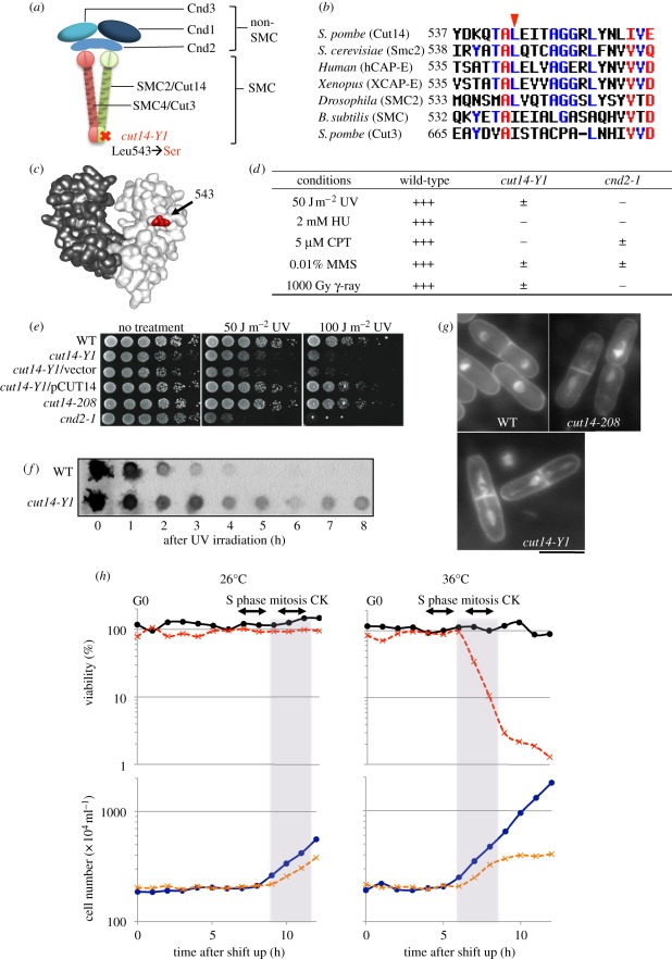
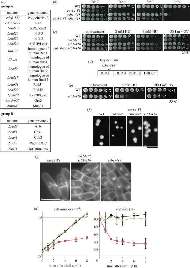
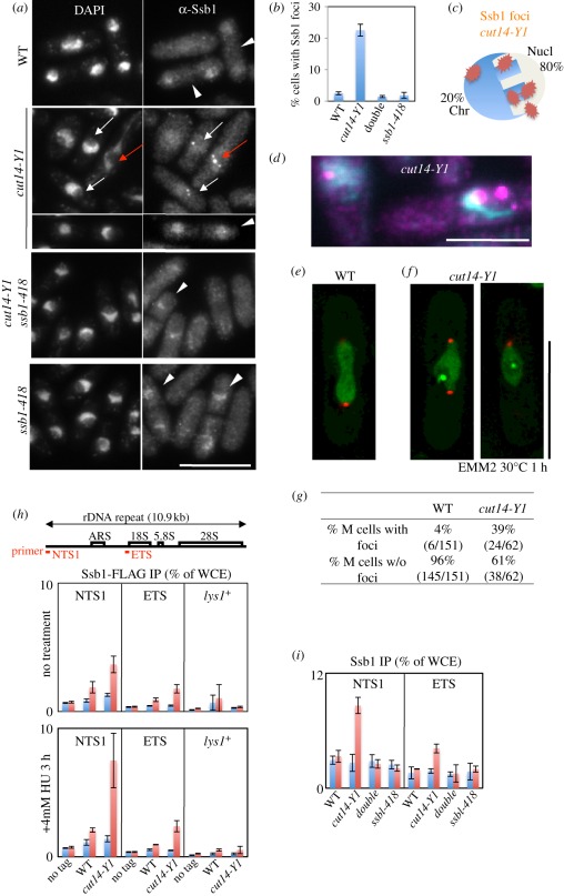
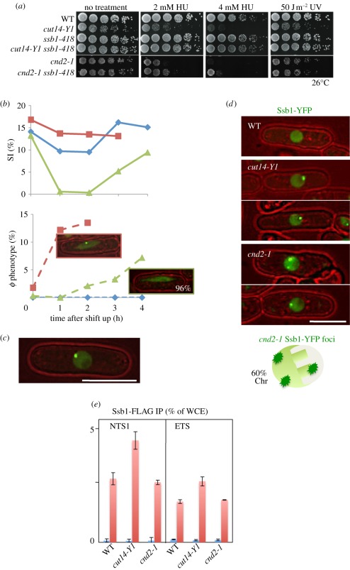
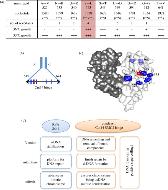
Condensin is required for chromosome dynamics and diverse DNA metabolism. How condensin works, however, is not well understood. Condensin contains two structural maintenance of chromosomes (SMC) subunits with the terminal globular domains connected to coiled-coil that is interrupted by the central hinge. Heterotrimeric non-SMC subunits regulate SMC. We identified a novel fission yeast SMC hinge mutant, cut14-Y1, which displayed defects in DNA damage repair and chromosome segregation. It contains an amino acid substitution at a conserved hinge residue of Cut14/SMC2, resulting in diminished DNA binding and annealing. A replication protein A mutant, ssb1-418, greatly alleviated the repair and mitotic defects of cut14-Y1. Ssb1 protein formed nucleolar foci in cut14-Y1 cells, but the number of foci was diminished in cut14-Y1 ssb1-418 double mutants. Consistent with the above results, Ssb1 protein bound to single-strand DNA was removed by condensin or the SMC dimer through DNA reannealing in vitro. Similarly, RNA hybridized to DNA may be removed by the SMC dimer. Thus, condensin may wind up DNA strands to unload chromosomal components after DNA repair and prior to mitosis. We show that 16 suppressor mutations of cut14-Y1 were all mapped within the hinge domain, which surrounded the original L543 mutation site.

Keywords: structural maintenance of chromosomes, DNA damage, mitosis, DNA metabolism, condensation.


Figures

Figure 1.
Figure 1.
Mutation site and phenotypes of condensin SMC mutant cut14-Y1. The identification of cut14-Y1: four mutant strains among 1300 ts strains examined exhibited condensation defects. Gene cloning, genetic analysis and gene sequencing established that the mutations resided in three distinct genes involved in chromosome condensation. Strain 393 was a DNA topoisomerase II top2 [26] mutant, strain 640 was a cut15 [27] (homologue of importin alpha) mutant and the remaining Y1 and 541 strains were cut14 [28,29] (SMC2 homologue) mutants. Since the cut14-Y1 strain was hypersensitive to DNA damage at 26°C (the permissive temperature), we examined whether the damage-sensitive phenotype was linked with the ts phenotype. Tetrad dissection demonstrated that the HU (hydroxyurea) and UV (ultraviolet) ray-sensitive phenotype co-segregated with the ts phenotype (electronic supplementary material, figure S1). (a) Heteropentameric condensin complex. The cut14-Y1 allele consists of a L543S substitution in the hinge. (b) Amino acid sequences of the SMC hinge that surround the mutation site (red arrowhead). (c) The mutation site (red) is shown within the three-dimensional structure of the mouse hinge domain [30]. (d) Summary of the DNA damage phenotypes of cut14-Y1 together with the previously reported response of cnd2-1 [31]. +++, normal growth; ±, very slow growth; −, no growth. (e) Wild-type (WT), cut14-Y1 and other strains were spot tested after UV irradiation at 26°C. (f) After UV irradiation (100 J m−2) at 26°C, extracts of the WT and cut14-Y1 cells harvested at intervals were immunoblotted using anti-thymine dimer antibodies. (g) The mitotic segregation defect of cut14-Y1 and cut14-208. DAPI was used to stain DNA. Scale bar, 10 µm. (h) WT and cut14-Y1 cells were first arrested at the pre-replicative G0 phase in nitrogen-deficient medium (EMM2-N) [32] at 26°C for 24 h, and then shifted to a nitrogen-replenished medium (EMM2) at 26°C (left) or at 36°C (right) for 12 h to measure cell viability (plated at 26°C) and cell number. The timing of S phase, mitosis and cytokinesis (CK) were determined by FACScan and DAPI-staining, respectively. Aliquots of the cultures were taken at 1 h intervals after replenishment, and 300 cells of each genotype were plated on three YPD plates for each time point. The plates were incubated at 26°C for 5 days, and the colony numbers were counted. Circles with solid line, WT; crosses with dashed line, cut14-Y1.
Figure 2.
Figure 2.
cut14-Y1 defects are additive with many DNA metabolic and checkpoint mutants, and are rescued by ssb1-418. (a) Strains crossed with cut14-Y1 to obtain the double mutants are shown. Crosses with group A (15 strains) did not yield viable double mutants, while those with group B (5 strains) produced double mutants with additive defects. Crosses with group C (14 strains) produced double mutants that did not display any additive defect (see also electronic supplementary material, figure S3). Group A included mutants that are involved with the 9-1-1 complex (rad9, rad1 and hus1), double strand break repair (rhp51, rad22, mus81 and ku70), replication (cdc6, cdc22 and orc5), 14-3-3 (rad24 and rad25), ssDNA nuclease (rad13) and DNA damage checkpoint (rad17 and rad26), many of which interact with single-stranded (ss) DNA or ssDNA-associated RPA [36,37]. Group B included five DNA checkpoint mutants (rad3, chk1, cds1, crb2 [38] and swi1 [39]), suggesting that the hinge of Cut14 might have a checkpoint function. Most of the group C mutants were related to replication, cell cycle and mitosis, although two were involved in DNA repair (uvde and rqh1 [40]). (b) ssb1-418 showed a striking synthetic rescue of the cut14-Y1 ts phenotype at 30°C and 33°C. (c) ssb1-418 also rescued the 2−4 mM HU and 50 J m−2 UV sensitivity of cut14-Y1 at 26°C. (d) The ssb1-418 strain contains a G78E amino acid substitution in the DBD F. (e) Single ssb1-418 mutants were sensitive to HU and UV at the semi-permissive temperature (33°C). (f) The colony formation of single and double mutants was examined at 26°C. Scale bar, 5 mm. The colony size of cut14-Y1 single mutants was smaller than that of the cut14-Y1 ssb1-418 double mutant at 26°C. The doubling time of single cut14-Y1 was 4.5 h at 26°C, while that of the double mutant and the WT was 3.5 h at 26°C. (g,h) In YPD liquid medium at 33°C, the cut14-Y1 single mutant lost viability and displayed frequent mitotic defects, while the double mutant and ssb1-418 grew normally. The asynchronous cultures of cut14-Y1, ssb1-418 and the double mutant in the YPD liquid medium were shifted from 26°C to 30°C. The cell number (per millilitre) was counted by the cell counter. The viability was measured at each time point by plating 300 cells spread on YPD plates, incubated at 26°C for 5 days, and resulting colonies were counted. Scale bar, 10 µm. (h) Black diamonds, ssb1-418; red squares, cut14-Y1; green triangles, cut14-Y1 ssb1-418.
Figure 3.
Figure 3.
Intense Ssb1-YFP foci formed in the nucleolus of cut14-Y1, but not in the double mutant. (a) Intense nuclear foci were detected by anti-Ssb1 antibody (DNA stained by DAPI). Immunofluorescence micrographs are shown for WT, single cut14-Y1, ssb1-418 and the double mutant cells that were shifted from 26°C to 30°C for 1 h. The Ssb1 foci were observed in both mitotic (red arrows) and interphase (white arrows) cut14-Y1 cells. Such foci were scarce in WT, ssb1-418 and cut14-Y1 ssb1-418 (double mutant) cells. The nuclear Ssb1 signals were clearly observed in S phase cells (white arrowheads). The S phase occurs in binucleated septated cells, allowing S phase cells to be easily distinguished. In those S phase cells, nuclear accumulation of Ssb1 appeared as punctate signals, distinct from the foci observed in cut14-Y1. Scale bar, 10 µm. (b) Quantitative data (percentage cells with Ssb1 foci) are shown. Three hundred cells were observed for each strain. (c) Anti-Ssb1 antibodies revealed that the intense foci were mostly (approx. 80%) located in the nucleolar region of the cut14-Y1 single mutant. The S. pombe interphase nucleus consists of the hemispherical chromatin region (Chr) and the remaining nucleolar (Nucl) region [43]. (d) The enlarged, merged micrograph of cut14-Y1 cells. Blue, DAPI; purple, Ssb1. Scale bar, 5 µm. (e,f) Live cell images of WT (e) and cut14-Y1 mutant (f) cells that express both Ssb1-YFP (green) and Sad1-mCherry (SPB, red). Scale bar, 10 µm. See electronic supplementary material, movies S1–S4. (g) The frequency (%) of cells showing Ssb1-YFP foci in WT and cut14-Y1 mitotic mutant cells were determined after the shift to 30°C (the restrictive temperature) from 26°C for 1 h in EMM2. (h) Chromatin immunoprecipitation (ChIP) experiment using anti-FLAG antibodies. The ssb1+ gene was chromosomally tagged with FLAG in the WT and cut14-Y1 strains, and expressed under the native promoter at 26°C in the absence (upper panel) or presence (lower panel) of 4 mM HU for 3 h. Two rDNA probes and one negative control (lys1+) probe were used. Blue and red columns indicate ChIP without and with antibodies against FLAG. An untagged (no tag) strain was used as the negative control. (i) ChIP experiment using anti-Ssb1 antibodies for the four strains cultured at 26°C. (h,i) Blue bars, −antibodies; red bars, +antibodies.
Figure 4.
Figure 4.
The checkpoint response and nuclear localization of Ssb1-YFP differ in cnd2-1 and cut14-Y1. (a) Whereas the DNA damage sensitivities of cut14-Y1 were greatly rescued by ssb1-418 mutation, those of cnd2-1 were not. (b) WT, cut14-Y1 and cnd2-1 were cultured at 36°C for 0–4 h, and the percentage septation index (SI) and the number of cells displaying aberrant chromosome (φ phenotype) were measured. The frequency of aberrant mitotic chromosomes sharply increased in cut14-Y1 (blue diamonds with solid line, WT; red squares with solid line, cut14-Y1; green triangles with solid line, cnd2-1; top), while the appearance of such mitotic cells was delayed in cnd2-1 (blue diamonds with dashed line, WT; red squares with dashed line, cut14-Y1; green triangles with dashed line, cnd2-1; bottom). Notably, the aberrant mitotic chromosomes in cnd2-1 did not contain Ssb1 foci (96%). (c) The intense foci of Ssb1-YFP were observed in cnd2-1 cells. Note that the YFP dot (the focus) is located in the nuclear periphery chromatin region. (d) Distinct nuclear localization of the Ssb1-YFP signals in cnd2-1 and cut14-Y1. The WT cell nucleus did not show the foci of Ssb1-YFP. Two cut14-Y1 cells display the intense Ssb1-YFP foci, which are located in the nucleolar region. Two cnd2-1 cells also show the intense Ssb1-YFP foci, which are located in the non-nucleolar nuclear chromatin region. Sixty per cent of cells examined showed the nuclear chromatin localization of Ssb1-YFP foci in cnd2-1 mutant cells. (e) ChIP experiment of Ssb1-FLAG for the rDNA probe NTS1 using WT, cut14-Y1 and cnd2-1 strains. The procedures are the same employed in figure 3h. Blue bars, −antibodies; red bars, +antibodies.
Figure 5.
Figure 5.
Interaction of isolated condensin and SMC dimer with different DNAs. (a) SDS-PAGE patterns of holocondensin (Cut3-Cut14-Cnd1-Cnd2-Cnd3), the SMC dimer (Cut3-Cut14) and the non-SMC trimer (Cnd1-Cnd2-Cnd3), together with single Cut3 and Cut14 as controls, stained with Coomasie brilliant blue. The procedures of isolation were previously described, and the degree of purity for these preparations was similar to those previously reported [20,21]. The Cut14 and Cnd1 overlap, and the Cnd2 band is diffuse and less intense than the other non-SMC subunits, probably owing to phosphorylation and/or degradation [9]. Limited proteolysis of Cut3 has been reported [20]. (b) Condensin and SMC dimer were incubated with a mixture of ssDNA and dsDNA, then analysed on a 10% non-denaturing acrylamide gel in the absence of SDS. DNA used was tagged with fluorescent FITC. (c) WT and mutant SMC dimer were incubated with M13 ssDNA with or without the pre-heat treatment at 42°C for 10 min, then analysed on a 0.7% native agarose gel in the absence of SDS. The mutant dimer was obtained by simultaneous overexpression of Cut3 and Cut14-Y1, and purified by affinity chromatography, stained with SYBR Gold. (d) WT and mutant SMC dimers were incubated with hdDNA with or without pre-heat treatment of the SMC dimers (see text), stained with ethidium bromide.
Figure 6.
Figure 6.
Condensin SMC-mediated elimination of RPA from hdDNA. (a) SMC dimer promotes reannealing of RPA-coated hdDNA. Lanes 1,2: control ds and hdDNA; 3–5: naked hdDNA (heat denatured and then rapidly cooled) was incubated with or without SMC for 0, 3 or 10 min; 6–9: hdDNA pre-coated with RPA was further incubated with (lanes 6–8) or without (lane 9) the SMC dimers. After incubation, samples were analysed on a 0.7% native agarose gel (without SDS). (b) Holocondensin also produced dsDNA from RPA-coated hdDNA. Native agarose gel was used. (c) hdDNA incubated with RPA complex was analysed in the absence or presence of SDS. See text. (d) Lanes 1,2: hdDNA incubated alone for 0 or 30 min; 3: dsDNA; 4–9: hdDNA pre-coated with SSB for 5 min at 30°C, and further incubated for 30 min without (lanes 4,5) or with SMC for 0–30 min (lanes 6–9). The reaction mixtures were analysed by native agarose gel electrophoresis. (e) AFM images hdDNA (top left), dsDNA (bottom left), hdDNA coated with SSB (middle). SMC was added and incubated with SSB-coated hdDNA for 30 min (right). (f) AFM images of hdDNA coated with S. pombe RPA (left); SMC dimer was added and incubated with RPA-coated hdDNA for 30 min (right). (g) Condensin and SMC dimer binding to RNA that was made in electronic supplementary material, figure S5. The samples were analysed using a 4% native agarose (NuSieve) gel in the absence of SDS. (h) (left) The mixture of hdDNA and DNA–RNA hybrid was digested with DNase I or RNase H. The hybrid band was selectively digested with RNase H. (right) Condensin and SMC dimers (0–100 nM) were incubated with the mixture, and SDS was used to stop the reactions. The samples were analysed using a 0.7% agarose gel. Staining with (a–d,h) ethidium bromide and (g) SYBR Gold.
Figure 7.
Figure 7.
Interaction of mutant RPA with DNA and the mutant SMC dimer. (a,b) Interaction of WT and mutant RPA complexes with (a) short and (b) long ssDNA. The heterotrimeric RPA that contained the Ssb1-418 mutant protein was purified and mixed with (a) short 86 nt ssDNA and (b) long M13 ssDNA, followed by (a) native acrylamide and (b) native agarose gel electrophoresis (in the absence of SDS). Binding of the mutant RPA to short 86 nt ssDNA was greatly diminished, whereas the binding to M13 ssDNA was only slightly diminished. (c) WT and mutant RPA (80 nM) were bound to heat-denatured hdDNA for 5 min on ice, followed by the addition of WT and mutant SMC dimer-containing Cut14-Y1 (0, 25, 50 nM) for the reannealing reaction at 30°C for 30 min. Resulting reaction mixtures were analysed using 0.7% native agarose gels and stained with ethidium bromide. Diffuse bands represented hdDNA coated with RPA, which formed with the WT and mutant RPA. The ability of mutant SMC dimer (Cut14-Y1) for reannealing was diminished for hdDNA precoated with the WT RPA, whereas the reannealing went equally well when the mutant RPA previously coated hdDNA. Staining with (a) FITC, (b) SYBR Gold and (c) ethidium bromide.
Figure 8.
Figure 8.
The hinge of SMC2/Cut14 is a functional entity. (a) Mapping of pseudo-revertants of cut14-Y1 that formed colonies at 33°C. The four mutants are true revertants (S543L, red column) that formed the normal colonies at 36°C. See text. (b) The mutation sites are shown by the vertical lines. The hinge region is between the residues 519 and 641. cc, coiled-coil. (c) Location of the mutation sites in the three-dimensional structure of the mouse condensin hinge [30]. The amino acid residue number is adapted for the S. pombe Cut14. The original cut14-Y1 mutation site is indicated by the red colour, while the second suppressing mutation sites are shown by blue. The residues 594 and 641 situating behind are faded. (d) A diagram depicting the relationship between the condensin SMC Cut14 hinge and Ssb1. Condensin preferentially binds to ssDNA [30,34] and promotes annealing to complementary ssDNA in vitro, and appears to oppose the action of RPA. RPA acts as a platform for various proteins involved in DNA metabolism, such as damage repair and replication through ssDNA stabilization [–46]. The role of condensin in damage repair remains unclear, but we propose that it may be required for completing/exiting repair processes by removing RPA and forming dsDNA through reannealing.

References

    1. Wood AJ, Severson AF, Meyer BJ. 2010. Condensin and cohesin complexity: the expanding repertoire of functions. Nat. Rev. Genet. 11, 391–40410.1038/nrg2794 (doi:10.1038/nrg2794) - DOI - DOI - PMC - PubMed
    1. Hudson DF, Marshall KM, Earnshaw WC. 2009. Condensin: architect of mitotic chromosomes. Chromosome Res. 17, 131–14410.1007/s10577-008-9009-7 (doi:10.1007/s10577-008-9009-7) - DOI - DOI - PubMed
    1. Anderson DE, Losada A, Erickson HP, Hirano T. 2002. Condensin and cohesin display different arm conformations with characteristic hinge angles. J. Cell Biol. 156, 419–42410.1083/jcb.200111002 (doi:10.1083/jcb.200111002) - DOI - DOI - PMC - PubMed
    1. Yoshimura SH, Hizume K, Murakami A, Sutani T, Takeyasu K, Yanagida M. 2002. Condensin architecture and interaction with DNA: regulatory non-SMC subunits bind to the head of SMC heterodimer. Curr. Biol. 12, 508–51310.1016/S0960-9822(02)00719-4 (doi:10.1016/S0960-9822(02)00719-4) - DOI - DOI - PubMed
    1. Hirano M, Hirano T. 2002. Hinge-mediated dimerization of SMC protein is essential for its dynamic interaction with DNA. EMBO J. 21, 5733–574410.1093/emboj/cdf575 (doi:10.1093/emboj/cdf575) - DOI - DOI - PMC - PubMed

Publication types

MeSH terms