

Valvular heart diseases in the developing world: developmental biology takes center stage
- PMID: 22645860
- PMCID: PMC3694273
Valvular heart diseases in the developing world: developmental biology takes center stage
Abstract
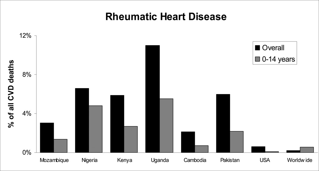
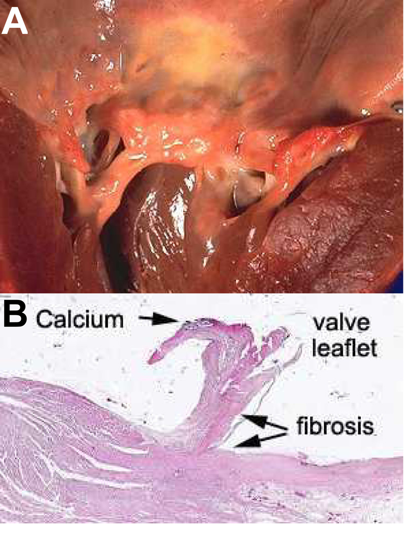
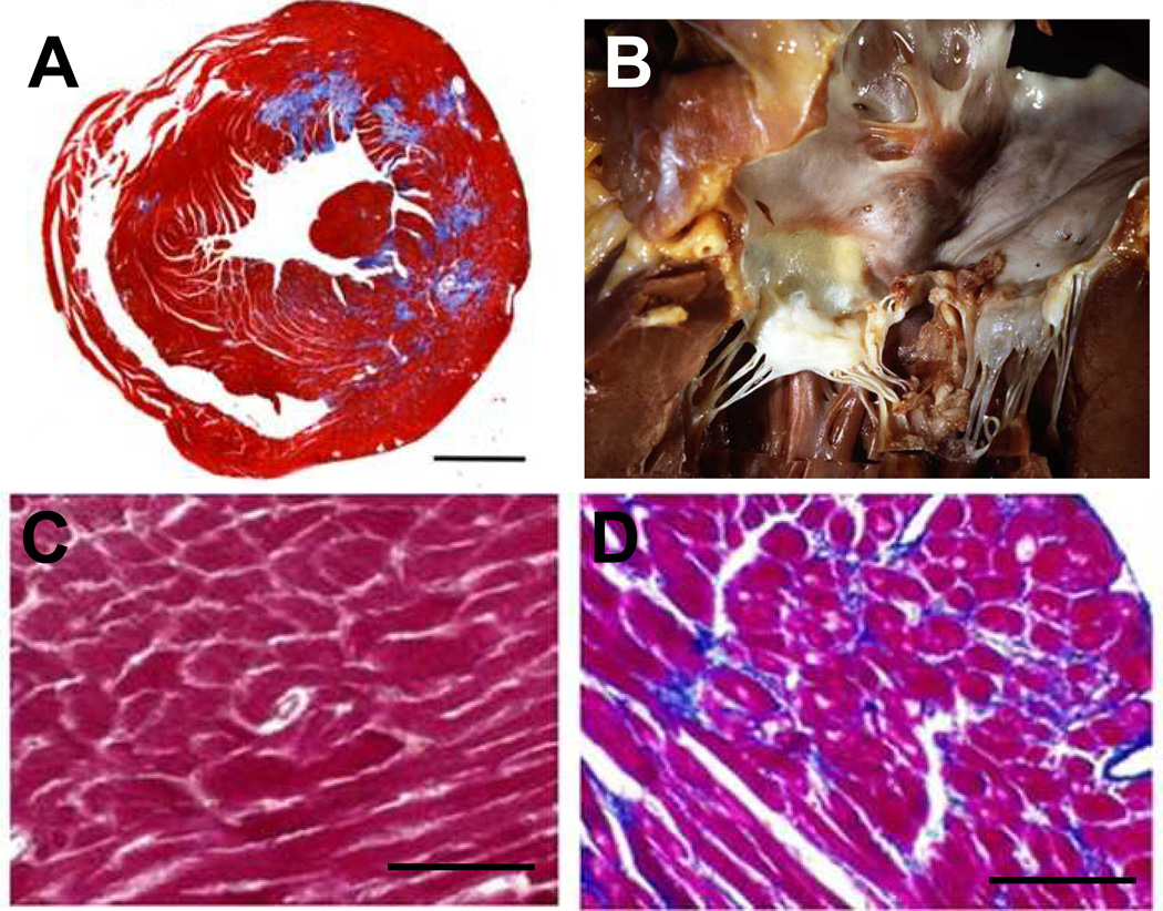
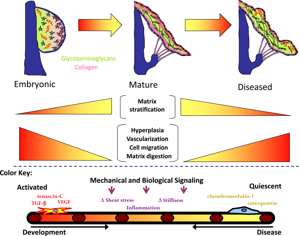
Heart valve disease is a significant and increasing global problem of which, in the developing world, the primary sufferers are the children and young adults regarded as the critical 'engine' of future economic growth. Yet, up to 10 times the current number of known sufferers remain undiagnosed in these countries. Among the most prevalent and neglected diseases are rheumatic heart disease and endomyocardial fibrosis. The etiologies of these diseases can be described in part as a dysregulation or reactivation of developmental biology pathways. Consequently, connecting mechanisms of valvulogenesis and disease etiology may represent an excellent strategy to identify therapeutic targets. These local diseases require local solutions tailored to local resources; therefore, collaboration with experienced research groups should be encouraged as a way of accelerating the creation of relevant knowledge, and its clinical translation.
Figures
References
-
- Yusuf S, Vaz M, Pais P. Tackling the challenge of cardiovascular disease burden in developing countries. Am Heart J. 2004;148:1–4. - PubMed
-
- Mathers C, Fat DM, Boerma J. The global burden of disease: 2004 update. WHO. 2008
-
- Guilherme L, Ramasawmy R, Kalil J. Rheumatic fever and rheumatic heart disease: Genetics and pathogenesis. Scand J Immunol. 2007;66:199–207. - PubMed
Publication types
MeSH terms
Grants and funding
LinkOut - more resources
Full Text Sources