. 2012 Jun 26;51(25):5187-97.
doi: 10.1021/bi3000462. Epub 2012 Jun 12.

An iron-sulfur cluster loop motif in the Archaeoglobus fulgidus uracil-DNA glycosylase mediates efficient uracil recognition and removal

Affiliations

An iron-sulfur cluster loop motif in the Archaeoglobus fulgidus uracil-DNA glycosylase mediates efficient uracil recognition and removal

Lisa M Engstrom et al. Biochemistry. .

Abstract

The family 4 uracil-DNA glycosylase from the hyperthermophilic organism Archaeoglobus fulgidus (AFUDG) is responsible for the removal of uracil in DNA as the first step in the base excision repair (BER) pathway. AFUDG contains a large solvent-exposed peptide region containing an α helix and loop anchored on each end via ligation of two cysteine thiolates to a [4Fe-4S](2+) cluster. We propose that this region plays a similar role in DNA damage recognition as a smaller iron-sulfur cluster loop (FCL) motif in the structurally unrelated BER glycosylases MutY and Endonuclease III and therefore refer to this region as the "pseudo-FCL" in AFUDG. In order to evaluate the importance of this region, three positively charged residues (Arg 86, Arg 91, Lys 100) and the anchoring Cys residues (Cys 85, Cys 101) within this motif were replaced with alanine, and the effects of these replacements on uracil excision in single- and double-stranded DNA were evaluated. These results show that this region participates and allows for efficient recognition and excision of uracil within DNA. Notably, R86A AFUDG exhibited reduced activity for uracil removal only within double-stranded DNA, suggesting an importance in duplex disruption and extrusion of the base as part of the excision process. In addition, mutation of the [4Fe-4S](2+) cluster cysteine ligands at the ends of the pseudo-FCL to alanine reduced the uracil excision efficiency, suggesting the importance of anchoring the loop via coordination to the cluster. In contrast, K100A AFUDG exhibited enhanced uracil excision activity, providing evidence for the importance of the loop conformation and flexibility. Taken together, the results herein provide evidence that the pseudo-FCL motif is involved in DNA binding and catalysis, particularly in duplex DNA contexts. This work underscores the requirement of an ensemble of interactions, both distant and in proximity to the damaged site, for accurate and efficient uracil excision.


Figures

Figure 1
Figure 1
A view of the pseudo-FCL motif in TTB (A) and the FCL motif in B. stearothermophilus MutY (B) and E.coli EndoIII (C). From structure coordinates deposited in the Protein Database, accession codes 2DDG, 1RRS, and 1ORP. Residues that make up the pseudo-FCL and FCL motifs are highlighted in fuschia, with bound DNA in blue.
Figure 2
Figure 2
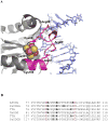
The pseudo-FCL motif in Family 4 and 5 UDGs. (A) Illustration of the close proximity of the pseudo-FCL motif (magenta) with duplex DNA (blue). The figure was generated by overlay of the structure coordinates of TTA (1UI0) and TTB co-crystallized with DNA (2DDG). Residue side chains that differed (Ile ✱Lys) were changed and numbered to correspond to those in AFUDG (see 2B for alignment). The positively charged residues in AFUDG that were replaced by alanine are shown on TTA structure in ball and stick representation. The [4Fe-4S] cluster is shown as spheres (Fe in orange, S in yellow). Note that only the side chains of the two Cys ligands that are part of the pseudo-FCL are shown. In addition, Cys 101 is coordinated to the iron that is behind the other three iron atoms and therefore is not visible. (B) Sequence alignments of the pseudo-FCL motif of various Family 4 and 5 UDGs. Conserved cysteine residues that ligate the [4Fe-4S]2+ cluster are highlighted in red, with positively charged residues within the pseudo-FCL motif highlighted in black.
Figure 3
Figure 3
Uracil excision from duplex substrates with TTB and AFUDG under multiple-turnover conditions. AFUDG exhibits fast substrate turnover resulting in depletion of substrate within the time-frame measured, while under similar conditions TTB displays biphasic kinetic behavior due to slow DNA product release. AFUDG (1 nM) and TTB (2 nM) incubated with 20 nM uracil-containing duplex DNA, incubated at 52 °C in 20 mM Tris-HCl, 30 mM NaCl, 1 mM EDTA.
Figure 4
Figure 4
Representative graph of binding titrations of AFUDG with RhX-product analog ds DNA using fluorescence anisotropy. Reaction conditions as follows: [DNA] = 500 nM, [NaCl] = 30 mM, reactions incubated at 25 °C. Active fraction was determined from an average of at least three independent titrations.
Figure 5
Figure 5
Active fraction of TTB determined from glycosylase activity and fluorescence anisotropy binding titrations. (A) Active fraction determined from burst amplitudes for MTO reactions under the following conditions: TTB (2 nM, 4 nM, 8 nM) incubated with 20 nM uracil-containing duplex DNA, incubated at 37 °C. (B) Anisotropy reactions were carried out as follows: [DNA] = 500 nM, [NaCl] = 30 mM, reactions incubated at 25 °C. Active fraction determined from an average of at least three independent titrations.
Figure 6
Figure 6
Multiple-turnover glycosylase activity of AFUDG. (A) Representative data for AFUDG catalyzed uracil excision from a double-stranded DNA containing a centrally located U:G mispair. (B) Representative data for AFUDG catalyzed uracil excision from a single-stranded DNA containing a centrally located uracil. For both single- and double-stranded experiments, reaction conditions were as follows: [E] = 1 nM, [DNA] = 10 – 200 nM, reactions incubated at 52 °C. The lines indicate the best fit to the Michaelis-Menten equation, and relevant kcat and Km parameters from at least three separate experiments are listed in Table 1.
Figure 7
Figure 7
Representative plots of single-turnover excision of uracil in ds DNA by WT and R86A AFUDG. Lines represent fitting to a single-exponential as described in the methods. At least three independent trials were used to determine values for k2 listed in Tables 1 and 2. For both single- and double-stranded experiments, reaction conditions were as follows: [E] ≥ 400 nM, [DNA] = 20 nM, reactions incubated at 52 °C in 25 mM Tris-HCl, 30 mM NaCl, 1 mM EDTA, pH 7.6.
Figure 8
Figure 8
A view of the pseudo-FCL motif in TTB (A) and the analogous loop in other members of the UDG superfamily, human UDG (B) and Herpes simplex virus UDG (C). Figures were prepared from coordinates deposited in the Protein Database, accession codes 2DDG, 1SSP, and 1LAU, respectively. The DNA is shown in blue. Residues that make up the pseudo-FCL and analogous motifs are highlighted in fuchsia with a conserved Arg residue within this loop motif in each enzyme represented in a ball and stick model. Human UDG Arg 276, which has previously been shown to be important for binding and uracil removal, is colored in orange in ball and stick representation. The uracil and phosphate of the DNA is shown in ball and stick and green.
Scheme I
Scheme I

References

    1. David SS, Williams SD. Chemistry of Glycosylases and Endonucleases Involved in Base-Excision Repair. Chem Rev. 1998;98:1221–1262. - PubMed
    1. Lindahl T. Instability and decay of the primary structure of DNA. Nature. 1993;362:709–715. - PubMed
    1. Tye BK, Nyman PO, Lehman IR, Hochhauser S, Weiss B. Transient accumulation of Okazaki fragments as a result of uracil incorporation into nascent DNA. Proc Natl Acad Sci U S A. 1977;74:154–157. - PMC - PubMed
    1. Pearl LH. Structure and function in the uracil-DNA glycosylase superfamily. Mutat Res. 2000;460:165–181. - PubMed
    1. Lee HW, Dominy BN, Cao W. A new family of deamination repair enzymes in the uracil DNA glycosylase superfamily. J Biol Chem - PMC - PubMed

Publication types

MeSH terms