Review
. 2012 Sep;167(2):324-52.
doi: 10.1111/j.1476-5381.2012.02057.x.

Alzheimer's disease, β-amyloid, glutamate, NMDA receptors and memantine--searching for the connections

Affiliations
Review

Alzheimer's disease, β-amyloid, glutamate, NMDA receptors and memantine--searching for the connections

Wojciech Danysz et al. Br J Pharmacol. 2012 Sep.

Abstract

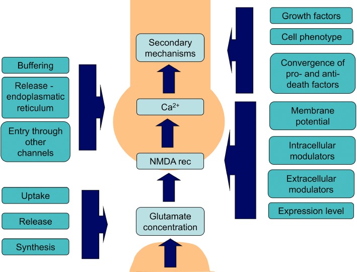
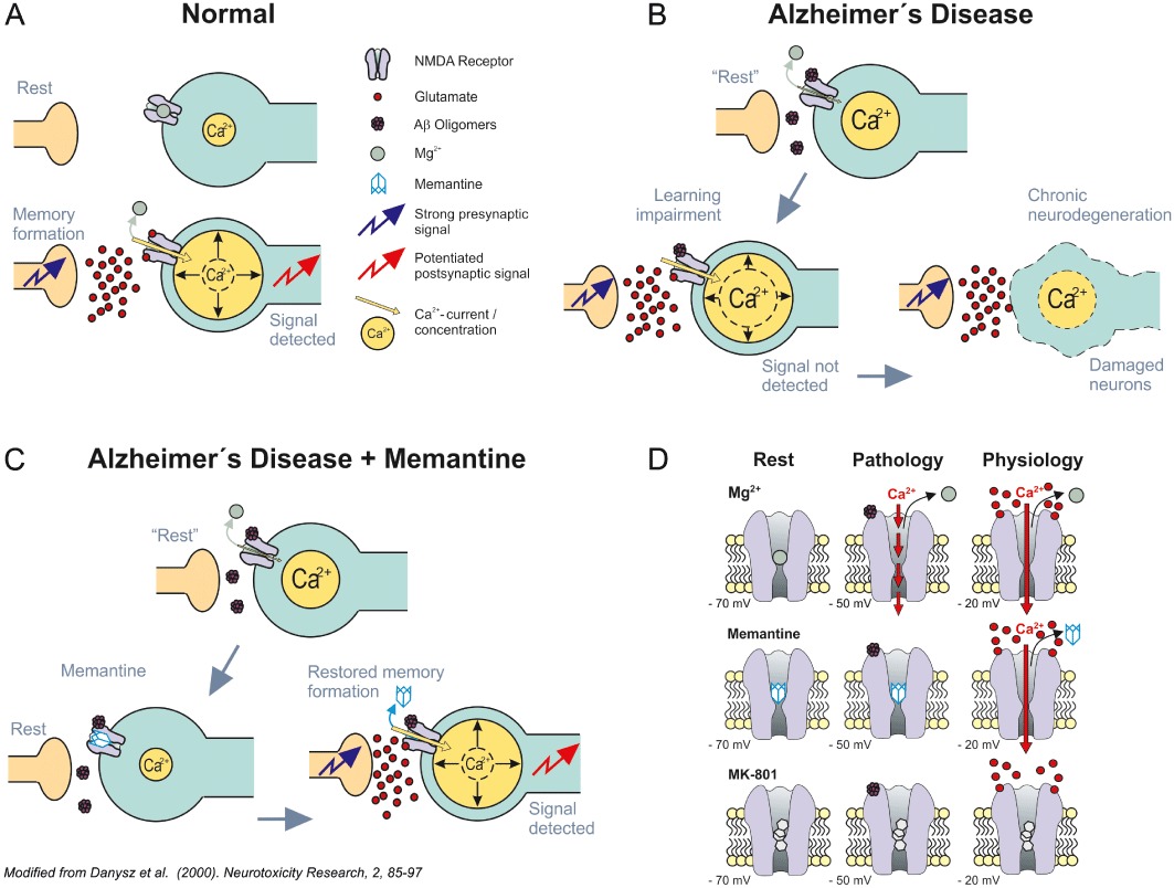
β-amyloid (Aβ) is widely accepted to be one of the major pathomechanisms underlying Alzheimer's disease (AD), although there is presently lively debate regarding the relative roles of particular species/forms of this peptide. Most recent evidence indicates that soluble oligomers rather than plaques are the major cause of synaptic dysfunction and ultimately neurodegeneration. Soluble oligomeric Aβ has been shown to interact with several proteins, for example glutamatergic receptors of the NMDA type and proteins responsible for maintaining glutamate homeostasis such as uptake and release. As NMDA receptors are critically involved in neuronal plasticity including learning and memory, we felt that it would be valuable to provide an up to date review of the evidence connecting Aβ to these receptors and related neuronal plasticity. Strong support for the clinical relevance of such interactions is provided by the NMDA receptor antagonist memantine. This substance is the only NMDA receptor antagonist used clinically in the treatment of AD and therefore offers an excellent tool to facilitate translational extrapolations from in vitro studies through in vivo animal experiments to its ultimate clinical utility.


Figures

Figure 1
Figure 1
Schematic illustrating the factors directly involved in normal physiological NMDA receptor-mediated synaptic transmission/plasticity and associated processes/factors that can modulate such NMDA receptor activation/transmission both under physiological, but more importantly also under disturbed pathological conditions. For simplification, the roles of other receptors (e.g. AMPA) and feedback inhibition in synaptic plasticity have been omitted from this cartoon (see also Figure 2A). The points where such secondary factors interact with this signalling cascade are indicated by the vertical blue boxes with associated arrows pointing to the light blue boxes.
Figure 2
Figure 2
(A) Under normal physiological conditions, synaptic plasticity/learning depends on the detection of a relevant (sufficiently strong) synaptic signal over background noise (here referring to transient high vs. prolonged moderate intracellular Ca2+ levels), resulting in a sufficient signal-to-noise ratio. Intracellular Ca2+ concentrations at any one time point are represented by different sizes of the yellow Ca2+ containing circles. Jagged blue arrow indicates arrival of a presynaptic signal. Jagged red arrow indicates detection of the postsynaptic signal. Mg2+ and glutamate are illustrated by large green and small red circles respectively. For simplification, the roles of other receptors (e.g. AMPA) and feedback inhibition have been omitted from this cartoon (please refer to figure 3 in Parsons et al., 2007 for a more detailed depiction of the processes believed to underlie such synaptic plasticity). (B) The signal-to-noise ratio hypothesis assumes that in Alzheimer's disease (AD), due to a tonic over activation of NMDA receptors by, for example soluble β-amyloid oligomers, Mg2+ is no longer effective enough to play its ‘filtering’ function. In turn, synaptic noise rises, impairing detection of the relevant synaptic signal required for learning/plasticity. The light blue straight arrows indicate the proposed course of events (i.e. first symptomatic disturbance of synaptic plasticity) followed by synaptotoxicity and ultimately neuronal death. Soluble β-amyloid oligomers represented as mauve aggregates of small circles – here binding directly to NMDA receptors for simplification, but probably interacting more directly with anchoring protein complexes and thereby affecting the function of their associated proteins such as NMDA receptors. Other symbols have the same meaning as in panel A. (C) Schematic illustrating memantine's proposed MoA in AD based on the signal-to-noise hypothesis. Memantine is able to serve as a more effective filter than Mg2+, blocking pathological ‘noise’ at glutamatergic synapses and thereby allowing detection of the relevant synaptic signal. Synaptic plasticity is restored and synaptotoxicity/ultimate neuronal death is prevented by the same MoA. Memantine illustrated as a simple light blue adamantane cage. For other aspects, see legends to panels A and B. Modified from Danysz et al. (2000). (D) Schematic illustrating the hypothesis explaining how the fast unblocking kinetics of memantine allow this voltage-dependent compound to differentiate between the physiological and pathological activation of NMDA receptors. Under resting therapeutic conditions [i.e. in their continuing presence at −70 mV (left), Mg2+ (top), memantine (middle) and MK-801 (bottom)], all occupy the NMDA receptor channel. Both Mg2+ and memantine are able to leave the NMDA receptor channel upon strong synaptic depolarization (−20 mV, right) due to their pronounced voltage dependency and rapid unblocking kinetics, whereas the slow, potent blocker MK-801 remains trapped. However, memantine – in contrasts to Mg2+– does not leave the channel so easily upon moderate prolonged depolarization during chronic excitotoxic insults caused by soluble β-amyloid oligomers tonically activating NMDA receptors (−50 mV, centre). Transient strong and prolonged moderate Ca2+ influx illustrated by the full and dashed red arrows respectively. Modified after Kornhuber and Weller (1997) and Parsons et al. (1999).

References

    1. Abbott JJ, Howlett DR, Francis PT, Williams RJ. Abeta(1-42) modulation of Akt phosphorylation via alpha7 nAChR and NMDA receptors. Neurobiol Aging. 2007;29:992–1001. - PubMed
    1. Abe K, Misawa M. Amyloid beta protein enhances the clearance of extracellular L-glutamate by cultured rat cortical astrocytes. Neurosci Res. 2003;45:25–31. - PubMed
    1. Abramov E, Dolev I, Fogel H, Ciccotosto GD, Ruff E, Slutsky I. Amyloid-beta as a positive endogenous regulator of release probability at hippocampal synapses. Nat Neurosci. 2009;12:1567–1576. - PubMed
    1. Alberdi E, Sanchez-Gomez MV, Cavaliere F, Perez-Samartin A, Zugaza JL, Trullas R, et al. Amyloid beta oligomers induce Ca2+ dysregulation and neuronal death through activation of ionotropic glutamate receptors. Cell Calcium. 2010;47:264–272. - PubMed
    1. Albrecht M, Rammes G, Parsons CG. 2008. Memantine reverses β-amyloid oligomers-induced deficits in long term potentiation (LTP) in murine hippocampal slices. 38th Society for Neuroscience Annual Meeting. Washington, USA. 15-11-2008. Society for Neuroscience Abstracts. 34, #829.21.

MeSH terms