. 2012 Oct;14(10):1554-67.
doi: 10.1111/j.1462-5822.2012.01820.x. Epub 2012 Jun 19.

Chlamydial infection induces host cytokinesis failure at abscission

Affiliations

Chlamydial infection induces host cytokinesis failure at abscission

Heather M Brown et al. Cell Microbiol. 2012 Oct.

Abstract

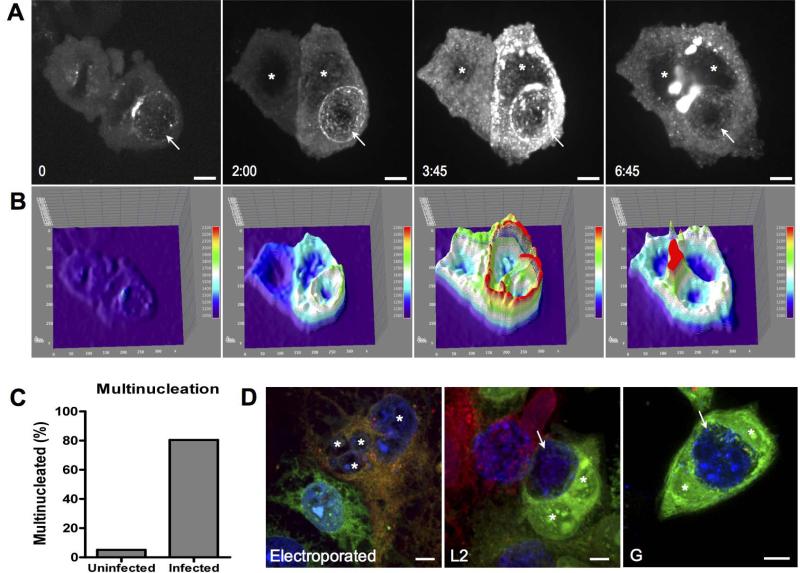
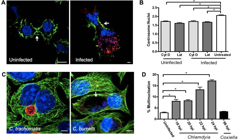
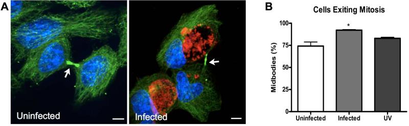
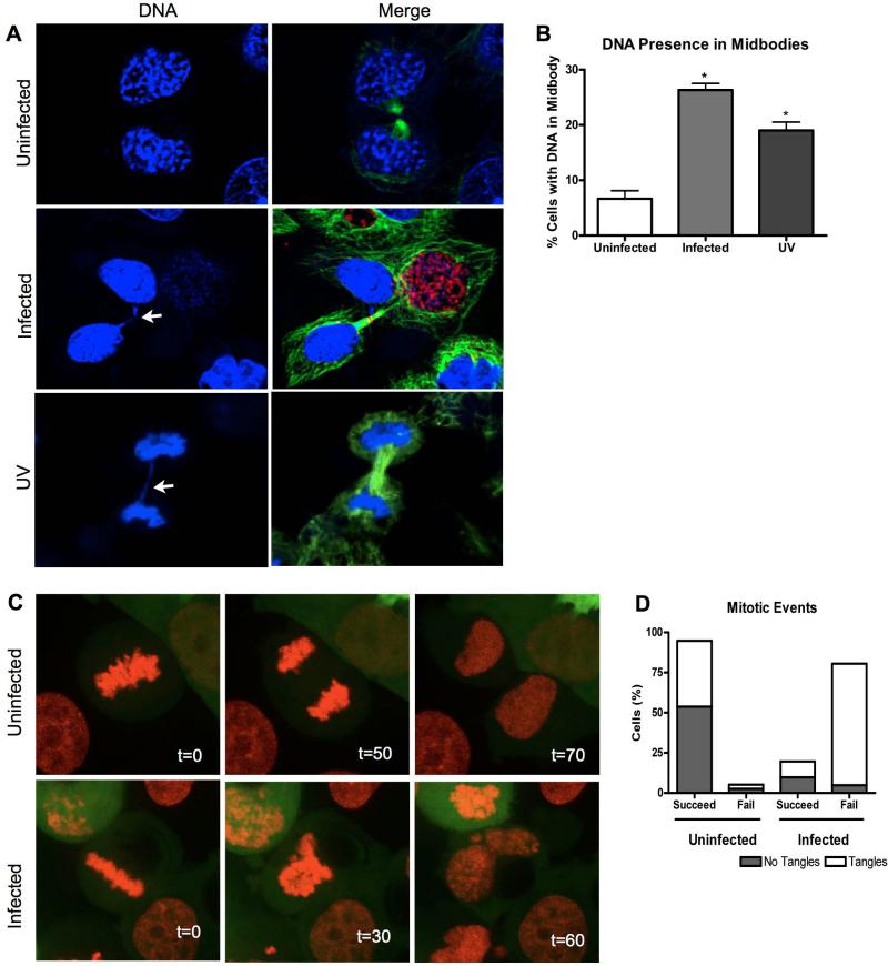
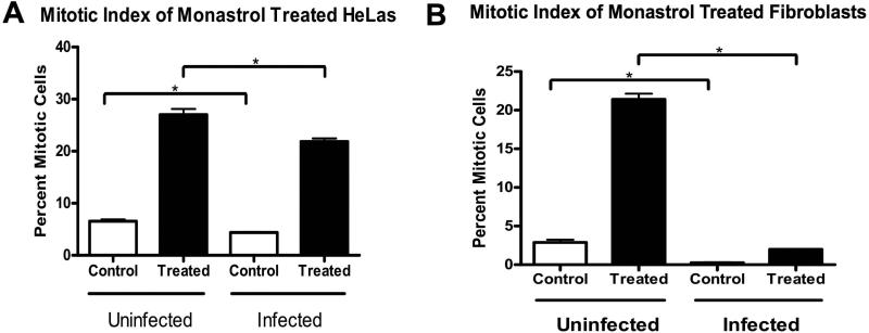
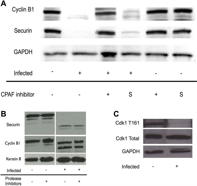
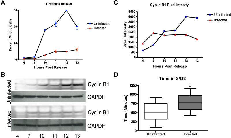
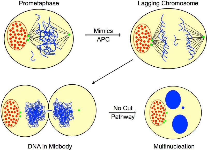
Chlamydia trachomatis is an obligate intracellular bacteria and the infectious agent responsible for the sexually transmitted disease Chlamydia. Infection with Chlamydia can lead to serious health sequelae such as pelvic inflammatory disease and reproductive tract scarring contributing to infertility and ectopic pregnancies. Additionally, chlamydial infections have been epidemiologically linked to cervical cancer in patients with a prior human papilomavirus (HPV) infection. Chlamydial infection of cultured cells causes multinucleation, a potential pathway for chromosomal instability. Two mechanisms that are known to initiate multinucleation are cell fusion and cytokinesis failure. This study demonstrates that multinucleation of the host cell by Chlamydia is entirely due to cytokinesis failure. Moreover, cytokinesis failure is due in part to the chlamydial effector CPAF acting as an anaphase promoting complex mimic causing cells to exit mitosis with unaligned and unattached chromosomes. These lagging and missegregated chromosomes inhibit cytokinesis by blocking abscission, the final stage of cytokinesis.


Figures

Figure 1
Figure 1. Multinucleation of host cells is due to cytokinesis failure and not cell fusion
[A-B] HeLa cells were transfected with GFP-Bicaudal D1 and then infectedC. trachomatis L2 for 24 hours. The cells were then imaged every 15 minutes for 18 hours. [A]. Images are time points from live cell imaging. Time 0 is started at telophase of the cell. Stars mark nuclei while the arrow points to the inclusion. [B] GFP signal was graphed with an intensity plot to allow visual comparison of differing intensities between forming daughter cells. [C] Live cell imaging was performed on Chlamydia infected HeLa cells. Beginning at 20 hpi cells were imaged every 10 minutes for 24 hours. Uninfected mitotic cells resulted in multinucleation in 5.1% of the cells, while infected mitotic cells became multinucleated 80.5%. n=39 and n=41, respectively. [D] Two different populations of HeLa cells were stained with Cytotracker Red or Green. Cells were co-plated and either electroporated or infected with C. trachomatis L2 or G. Electroporation caused fusion as shown by the yellow multinucleated cell. Neither serovars of C. trachomatis induced fusion. 1000 multinucleated cells were counted and all multinucleated cells were due to cytokinesis failure.
Figure 2
Figure 2. Chlamydia does not sterically hinder cytokinesis
[A] HeLa cells were infected for 30 hours then fixed and imaged using immunofluorescence. Cells were stained with either phalloidin (green), Draq 5 for DNA (blue) and human serum for Chlamydia (red). The actin ring was able to properly form in Chlamydia infected cells. Arrows point to the actin ring. [B] HeLa cells were treated with cytochalasin D or latrunculin A and the centrosome to nuclei ratio was evaluated using anti-Ɣ-tubulin and Draq5. Uninfected cells and infected cells treated with cytochalasin D did not show a significant difference in centrosome to nuclei ratio, having a ratio of 1.7 ± 0.1 and 1.7 ± 0.1 respectively. Similarly, when cells were treated with latrunculin A, uninfected cells had a centrosome to nuclei ratio of 1.6 ± 0.1, while infected cells had a ratio of 1.7 ± 0.1 Untreated infected cells showed a significant increase with an average of 2.1 ± 0.1 centrosomes per cell p = 0.001. N= 3 experiments with at least 150 nuclei per experiment. [C] Cells were infected with Chlamydia for 18 hours or Coxiella for 96 hours, then fixed. Cells were stained with anti-β-tubulin (green), human serum (red), and Draq5 (blue). The arrow indicates the location of the CCV. [D] Cells were infected with Chlamydia or Coxiella for the indicated amounts of time and percent multinucleation of the population of infected cells were counted. Uninfected HeLa had a multinucleation rate of 2.9 ± 0.3% while infection increased multinucleation with time. The infected rates were 8.02 ± 0.81%, 8.26 ± 0.50%, 13.2 ± 1.3%, 17.2 ± 0.5% for 18, 20, 22, 24 hpi respectively. All chlamydial time points investigated demonstrated a significance of p < 0.005. Coxiella had a multinucleation rate of 3.4 ± 0.2%. N=3 experiments with at least 600 cells per experiment.
Figure 3
Figure 3. Chlamydial infected cells form midbodies
[A-B] HeLa cells were infected for 30 hours then fixed and imaged using immunofluorescence. Cells were stained with anti-β-tubulin (green), human serum (red), and Draq 5 (blue). Arrows indicate location of the midbody. [B] All cells that were post-metaphase in mitosis were evaluated for midbody formation. Midbodies were present in 74.2 ± 4.6 % of uninfected cells exiting mitosis, while this percentage rose to 92.2 ± 0.6% in cells infected with Chlamydia. A midbody was formed in 83.0 ± 1.2% of cells exiting mitosis that were exposed to UV light. Infected cells demonstrated a significant increase in midbody formation with a p=0.017. N= 3 experiments with at least 150 cells per experiment.
Figure 4
Figure 4. Chlamydia induces lagging chromosomes in infected cells
[A] HeLa cells were infected for 30 hours then fixed and imaged. Conversely, uninfected cells were exposed to UV light for 3 minutes, allowed to grow for 24 hours and then fixed. Cells were stained with anti-β-tubulin (green), human serum (red), and Draq5 (blue) then examined in late telophase. Arrows indicate DNA bridging. [B] Uninfected cells had DNA in the midbody 6.7 ± 1.5% of the time, while infected cells had DNA in the midbody 26.3 ± 1.2% of the time. When cells were exposed to UV light there was DNA present in the midbody 19.00± 1.5% of the time. N=3 experiments with at least 200 cells per experiment. A student t-test was performed with p<0.02. [C] Stable RFP-H2B HeLa cells were transfected with GFP and infected with L2 for 20 hours. Cells were then imaged every 10 minutes for 24 hours. Panels show metaphase at time 0, followed by anaphase second and finally telophase of uninfected and infected cells. [D] Cells were followed through mitosis with live cell imaging as shown in C. Cells were scored based on if they failed or succeeded in mitosis along with if DNA missegregation events were present. In uninfected cells, 94.9% of cells had successful mitosis, while only 19.5% of infected cells were successful. N=39 and N=41 cells, respectively.
Figure 5
Figure 5. Chlamydia infection causes the premature exit of mitosis
HeLa cells were fixed 30 hpi and stained with anti-β-tubulin (green), human serum (red), and Draq5 (blue). Mitotic cells were compared to the total number of cells after infection and/or monastrol treatment. N= 3 experiments with at least 1500 cells per experiment. [A] Uninfected HeLa cells had a mitotic index of 6.6 ± 0.3% which decreased to 4.4 ± 0.1% when infected, with a significance of p=0.003. When treated with monastrol 27.0 ± 1.1% of uninfected HeLa cells arrest in mitosis, but only 21.9 ± 0.6% of infected cells, p<0.01. [B] Fibroblasts had a mitotic index of 2.9 ± 0.4% and 0.3 ± 0.1% for uninfected and infected cellls, respectively, with a significance of p<0.002. Monastrol treated uninfected fibroblasts had a mitotic index of 21.4 ± 0.8%, while infected fibroblasts had a mitotic index of 1.9 ± 0.03%, with p<0.0001.
Figure 6
Figure 6. CPAF degrades cyclin B1 and securin
[A] A cell-free degradation was performed and analyzed by western blot. Cyclin B1 and securin were degraded by infected cell lysate. Degradation could be blocked with a small peptide inhibitor of CPAF. [B] Same experiment as A, but infected cell lysate was treated with a protease inhibitor cocktail. [C] Mitotic shake-offs were performed to harvest proteins from uninfected and infected cells. Western blots show that the level of Cdk1 is unchanged due to infection, but the active T161 form of Cdk1 is not present in infected cells.
Figure 7
Figure 7. Host cell entry into mitosis
[A-B] A double thymidine block and infection was performed on HeLa cells. Cells were released and samples were collected at time points indicated. [A] Cells were fixed and stained for anti-β-tubulin (green), human serum (red), and Draq5 (blue) and a mitotic index was performed. In uninfected cells, a significant increase in mitotic cells begins at 10 hours post release, with the peak of mitosis occurring 12 hours after release from thymidine. Infected cells begin entering mitosis at 10 hours as well, but are not tightly synchronized. [B] Western blot analysis was performed on uninfected and infected cell lysates. Cyclin B1 levels were evaluated for cells entering mitosis. [C] The cyclin B1 western blot was analyzed for the mean pixel intensity of the bands and graphed. [D] HeLa cells were transduced using the FUCCI system and infected with Chlamydia. Starting 18 hpi, the cells were imaged every 10 minutes for 18 hours. The duration of S/G2 was calculated from the time of Cdt1-RFP degradation to mitotic entry (nuclear membrane breakdown). Uninfected cells were in S/G2 for 507 ± 78.4 minutes while infected required 774.6 ± 68.7 minutes with N=11, 13 respectively. The duration was significantly different with p<0.02.
Figure 8
Figure 8. Model of chlamydial induced multinucleation
Chlamydial inclusions (red) secrete CPAF into the cytoplasm of the host cell (yellow). CPAF mimics the APC causing the cell to exit metaphase before microtubules (black) attach to the kinetochores of the chromosomes (blue). Furthermore, premature exit from metaphase does not allow supernumerary centrosomes (green) to cluster. This results in lagging chromosomes that become trapped in the midbody of the host cell. The host cell aborts abscission via the No Cut Pathway in order to protect the DNA. Lagging chromosomes can result in micronuclei.

Similar articles

Cited by

References

    1. Alzhanov DT, Weeks SK, Burnett JR, Rockey DD. Cytokinesis is blocked in mammalian cells transfected with Chlamydia trachomatis gene CT223. BMC Microbiol. 2009;9:2. - PMC - PubMed
    1. Anttila T, et al. Serotypes of Chlamydia trachomatis and risk for development of cervical squamous cell carcinoma. JAMA. 2001;285:47–51. - PubMed
    1. Baker DJ, Dawlaty MM, Galardy P, van Deursen JM. Mitotic regulation of the anaphase-promoting complex. Cell. Mol. Life Sci. 2007;64:589–600. - PMC - PubMed
    1. Balsara ZR, Misaghi S, Lafave JN, Starnbach MN. Chlamydia trachomatis infection induces cleavage of the mitotic cyclin B1. Infect. Immun. 2006;74:5602–5608. - PMC - PubMed
    1. Caballe A, Martin-Serrano J. ESCRT machinery and cytokinesis: the road to daughter cell separation. Traffic. 2011;12:1318–1326. - PubMed

Publication types