. 2012 Aug 1;303(3):R301-10.
doi: 10.1152/ajpregu.00114.2012. Epub 2012 May 30.

Role of small conductance calcium-activated potassium channels expressed in PVN in regulating sympathetic nerve activity and arterial blood pressure in rats

Affiliations

Role of small conductance calcium-activated potassium channels expressed in PVN in regulating sympathetic nerve activity and arterial blood pressure in rats

Le Gui et al. Am J Physiol Regul Integr Comp Physiol. .

Abstract

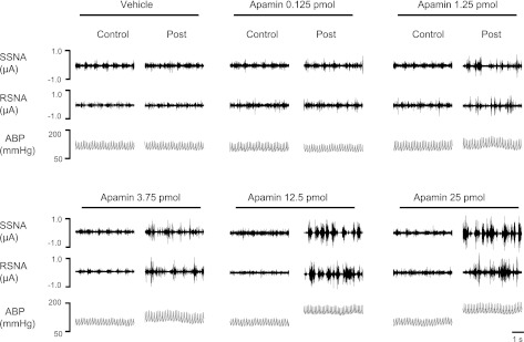
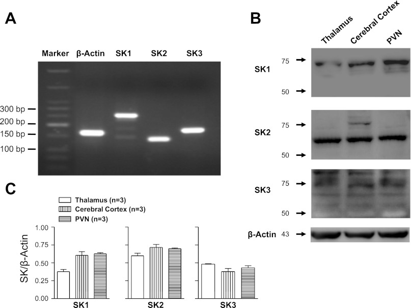
Small conductance Ca(2+)-activated K(+) (SK) channels regulate membrane properties of rostral ventrolateral medulla (RVLM) projecting hypothalamic paraventricular nucleus (PVN) neurons and inhibition of SK channels increases in vitro excitability. Here, we determined in vivo the role of PVN SK channels in regulating sympathetic nerve activity (SNA) and mean arterial pressure (MAP). In anesthetized rats, bilateral PVN microinjection of SK channel blocker with peptide apamin (0, 0.125, 1.25, 3.75, 12.5, and 25 pmol) increased splanchnic SNA (SSNA), renal SNA (RSNA), MAP, and heart rate (HR) in a dose-dependent manner. Maximum increases in SSNA, RSNA, MAP, and HR elicited by apamin (12.5 pmol, n = 7) were 330 ± 40% (P < 0.01), 271 ± 40% (P < 0.01), 29 ± 4 mmHg (P < 0.01), and 34 ± 9 beats/min (P < 0.01), respectively. PVN injection of the nonpeptide SK channel blocker UCL1684 (250 pmol, n = 7) significantly increased SSNA (P < 0.05), RSNA (P < 0.05), MAP (P < 0.05), and HR (P < 0.05). Neither apamin injected outside the PVN (12.5 pmol, n = 6) nor peripheral administration of the same dose of apamin (12.5 pmol, n = 5) evoked any significant changes in the recorded variables. PVN-injected SK channel enhancer 5,6-dichloro-1-ethyl-1,3-dihydro-2H-benzimidazol-2-one (DCEBIO, 5 nmol, n = 4) or N-cyclohexyl-N-[2-(3,5-dimethyl-pyrazol-1-yl)-6-methyl-4-pyrimidin]amine (CyPPA, 5 nmol, n = 6) did not significantly alter the SSNA, RSNA, MAP, and HR. Western blot and RT-PCR analysis of punched PVN tissue showed abundant expression of SK1-3 channels. We conclude that SK channels expressed in the PVN play an important role in the regulation of sympathetic outflow and cardiovascular function.


Figures

Fig. 1.
Fig. 1.
Schematic drawings of rat hypothalamus in coronal section. Shaded areas indicate regions of hypothalamus exposed to injected dye (A). The shape of each area was determined by tracing the outline of the dye observed on each section through the paraventricular nucleus (PVN). B: representative of a single injection (50 nl) within the PVN. AH, anterior hypothalamic area; 3V, third cerebral ventricle; RCh, retrochiasmatic area; MPO, medial preoptic nucleus; opt, optic tract; SOX, supraopticdecussation; StHy, striohypothalamic nucleus.
Fig. 2.
Fig. 2.
Representative traces show heart rate (HR), splanchnic sympathetic nerve activity (SNA) (SSNA), renal SNA (RSNA), and arterial blood pressure (ABP) responses to bilateral microinjections of the small conductance Ca2+-activated K+ (SK) channel blocker apamin (12.5 pmol). A: each injection (50 nl) of apamin (arrowhead) was completed over a period of ∼2 min. The interval between two injections was ∼2 min. Note that HR, SSNA, RSNA, and ABP were markedly increased. All tracings were recorded in the same animal. B: left, 5-s specimen trace of SSNA (top) and RSNA (bottom) before injection of apamin into the PVN. Right, 5-s specimen trace of SSNA (top) and RSNA (bottom) after microinjection of apamin into the PVN. The gap between B, left and right, is ∼100 min.
Fig. 3.
Fig. 3.
Representative traces show the dose-dependent responses of SSNA, RSNA, and ABP to bilateral microinjections of graded concentrations of apamin into the PVN (vehicle, 0.125, 1.25, 3.75, 12.5, and 25 pmol) in the anesthetized rats. A 50-nl injection of apamin was completed over a period of ∼2 min.
Fig. 4.
Fig. 4.
Summary data showing the changes in SSNA, RSNA, MAP, and HR in response to bilateral microinjections of varying doses of apamin (vehicle, 0.125, 1.25, 3.75, 12.5, and 25 pmol) into the PVN. Note that graded concentrations of apamin elicited a dose-dependent increase in these recorded variables. *P < 0.05 vs. vehicle; †P < 0.01 vs. vehicle; #P < 0.05 vs. 3.75 pmol group.
Fig. 5.
Fig. 5.
Representative traces show HR, SSNA, RSNA, and ABP responses to bilateral microinjections of nonpeptide SK channel blockade with UCL1684 (250 pmol). A: 50-nl injection of UCL1684 (arrowhead) was completed over a period of ∼2 min. The interval between two injections was ∼2 min. All tracings were recorded in the same animal. B: left, 5-s specimen traces of SSNA (top) and RSNA (bottom) before microinjection of UCL1684 into the PVN. Middle, 5-s specimen traces of SSNA (top) and RSNA (bottom) after microinjection of UCL1684 into the PVN. Right, 5-s specimen traces of SSNA (top) and RSNA (bottom) after UCL1684 washout.
Fig. 6.
Fig. 6.
Summary data showing changes in HR, SSNA, RSNA, and MAP in response to bilateral microinjections of vehicle (n = 4), UCL1684 (2.5 pmol, n = 5), and UCL1684 (250 pmol, n = 7). *P < 0.05 vs. vehicle. #P < 0.05 vs. 2.5 pmol group.
Fig. 7.
Fig. 7.
Representative traces show HR, SSNA, RSNA, and ABP responses to bilateral microinjection of the SK channel enhancer DCEBIO (5 nmol). A: 50-nl injection of DCEBIO (arrowhead) was completed over a period of ∼2 min. The interval between two injections was ∼2 min. All tracings were recorded in the same animal. B: left, 5-s specimen traces of SSNA (top) and RSNA (bottom) before microinjection of DCEBIO into the PVN. Right, 5-s specimen traces of SSNA (top) and RSNA (bottom) after microinjection of DCEBIO into the PVN. The gap between B, left and right, is ∼100 min.
Fig. 8.
Fig. 8.
Gene and protein expression of SK1, SK2, and SK3 in PVN. A: detection of Kcnn channel isoform mRNAs in PVN in adult male rats. The cDNA generated from PVN was analyzed by PCR amplification using Kcnn1-3-specific primers (see Table 1). B: Western blot analysis shows the expression of SK channels protein in thalamus, cerebral cortex, and hypothalamic PVN tissues from adult male rats. The protein expression of SK1 (59 kd), SK2 (64 kd), and SK3 (82 kd) was detected in PVN. C: summary data of SK channels expression in brain tissues (n = 3).

Similar articles

Cited by

References

    1. Adelman JP, Maylie J, Sah P. Small-conductance Ca2+-activated K+ channels: form and function. Annu Rev Physiol 74: 245–269, 2012 - PubMed
    1. Bains JS, Ferguson AV. Paraventricular nucleus neurons projecting to the spinal cord receive excitatory input from the subfornical organ. Am J Physiol Regul Integr Comp Physiol 268: R625–R633, 1995 - PubMed
    1. Benitez BA, Belalcazar HM, Anastasia A, Mamah DT, Zorumski CF, Masco DH, Herrera DG, de Erausquin GA. Functional reduction of SK3-mediated currents precedes AMPA-receptor-mediated excitotoxicity in dopaminergic neurons. Neuropharmacology 60: 1176–1186, 2011 - PMC - PubMed
    1. Biancardi VC, Son SJ, Sonner PM, Zheng H, Patel KP, Stern JE. Contribution of central nervous system endothelial nitric oxide synthase to neurohumoral activation in heart failure rats. Hypertension 58: 454–463, 2011 - PMC - PubMed
    1. Boudaba C, Schrader LA, Tasker JG. Physiological evidence for local excitatory synaptic circuits in the rat hypothalamus. J Neurophysiol 77: 3396–3400, 1997 - PubMed

Publication types

MeSH terms

Substances