Review
. 2012 Sep;93(Pt 9):1853-1868.
doi: 10.1099/vir.0.043182-0. Epub 2012 May 30.

Application of next-generation sequencing technologies in virology

Affiliations
Review

Application of next-generation sequencing technologies in virology

Alan D Radford et al. J Gen Virol. 2012 Sep.

Abstract

The progress of science is punctuated by the advent of revolutionary technologies that provide new ways and scales to formulate scientific questions and advance knowledge. Following on from electron microscopy, cell culture and PCR, next-generation sequencing is one of these methodologies that is now changing the way that we understand viruses, particularly in the areas of genome sequencing, evolution, ecology, discovery and transcriptomics. Possibilities for these methodologies are only limited by our scientific imagination and, to some extent, by their cost, which has restricted their use to relatively small numbers of samples. Challenges remain, including the storage and analysis of the large amounts of data generated. As the chemistries employed mature, costs will decrease. In addition, improved methods for analysis will become available, opening yet further applications in virology including routine diagnostic work on individuals, and new understanding of the interaction between viral and host transcriptomes. An exciting era of viral exploration has begun, and will set us new challenges to understand the role of newly discovered viral diversity in both disease and health.


Figures

Fig. 1.
Fig. 1.
Scale of sequencing information available in GenBank. (a) Percentage growth of sequences deposited on GenBank between August 2009 and August 2010. Sequences are grouped based on the GenBank classification of division, either by their higher taxonomy or the sequencing method employed (Benson et al., 2011). (b) The 20 most frequently sequenced viruses appearing on GenBank. Sequences were identified based on their organism name as described in each submission; PRRSV, porcine reproductive and respiratory syndrome virus. These data were compiled using eid2 (Anonymous, 2012).
Fig. 2.
Fig. 2.
Preparation of DNA. (a) Double-stranded template DNA is (b) randomly degraded, size-fractionated and ligated to adapters. (c) Alternatively, targeted amplification of the template can also be carried out by PCR.
Fig. 3.
Fig. 3.
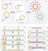
General principles of template amplification. (a–c) Emulsion PCR (Roche 454, SOLiD and Ion Torrent). (a) Adapters are used to capture single molecules of template onto microbeads by primer hybridization. (b) Beads are incorporated into a carefully controlled emulsion, in which each bubble constitutes a microreactor containing DNA template, primer and reagents for PCR. (c) Following amplification, each bead is coated with clonally amplified molecules. (d–h) Bridge amplification (Illumina). (d) Single-stranded template annealed to a glass plate by hybridization to a complementary primer. (e) The primer forms the basis for extension. (f) The free end of each single-stranded molecule can anneal to a second anchored primer in close spatial proximity, forming a ‘bridge’ that acts as a template for (g) a second round of amplification. This results in (h) four linear molecules. Stages (f)–(h) are essentially repeated to generate clonally amplified islands or clusters for subsequent sequencing. (i–m) Linear amplification (PacBio). (i) Template dsDNA. (j) Bound hairpin adapters create a single-stranded circular template. (k) Binding of a primer complementary to hairpin sequence. (l–m) Linear amplification and strand displacement create a single strand of DNA containing multiple copies of plus- and minus-strand sequences that serves as template for sequencing.
Fig. 4.
Fig. 4.
Methods of separating sequencing reactions. (a) Microbeads (Roche 454) or Ion Spheres (Ion Torrent) in microwells. (b) Clonally amplified beads bound to glass plate (SOLiD). (c) Amplified islands (Illumina). (d) Poly(A)-tagged library hybridized to plate (Helicos). (e) Amplified molecule captured by a single DNA polymerase molecule at the bottom of a microcell (PacBio).
Fig. 5.
Fig. 5.
Basic principles of sequencing chemistry. (a) Normal chain elongation leads to the release of (b) pyrophosphate (Roche 454) and (c) a hydrogen ion (Ion Torrent). (d) Structure of the ddNTP that forms the basis of Sanger sequencing. (e) Cartoon of the phospholinked fluorophore (PacBio). (f) Sequencing by ligation (SOLiD – see Fig. 6). (g) Cartoon of reversible chain terminators (Illumina and Helicos).
Fig. 6.
Fig. 6.
Basic principles underlying sequencing by ligation (synthesis) used by SOLiD.

References

    1. Ambrose H. E., Clewley J. P. (2006). Virus discovery by sequence-independent genome amplification. Rev Med Virol 16, 365–383 10.1002/rmv.515 - DOI - PMC - PubMed
    1. Anders S., Huber W. (2010). Differential expression analysis for sequence count data. Genome Biol 11, R106 10.1186/gb-2010-11-10-r106 - DOI - PMC - PubMed
    1. Anonymous (2003). Acute respiratory syndrome. China, Hong Kong Special Administrative Region of China, and Viet Nam. Wkly Epidemiol Rec 78, 73–74 - PubMed
    1. Anonymous (2012). ENHanCEd Infectious Diseases (eid2) database. http://www.zoonosis.ac.uk/EID2/, 20 April 2012.
    1. Baillie G. J., Galiano M., Agapow P. M., Myers R., Chiam R., Gall A., Palser A. L., Watson S. J., Hedge J. & other authors (2012). Evolutionary dynamics of local pandemic H1N1/2009 influenza virus lineages revealed by whole-genome analysis. J Virol 86, 11–18 10.1128/JVI.05347-11 - DOI - PMC - PubMed