. 2012 Jul 1;8(7):1045-57.
doi: 10.4161/auto.20159. Epub 2012 May 31.

Compromised autophagy by MIR30B benefits the intracellular survival of Helicobacter pylori

Affiliations

Compromised autophagy by MIR30B benefits the intracellular survival of Helicobacter pylori

Bin Tang et al. Autophagy. .

Abstract

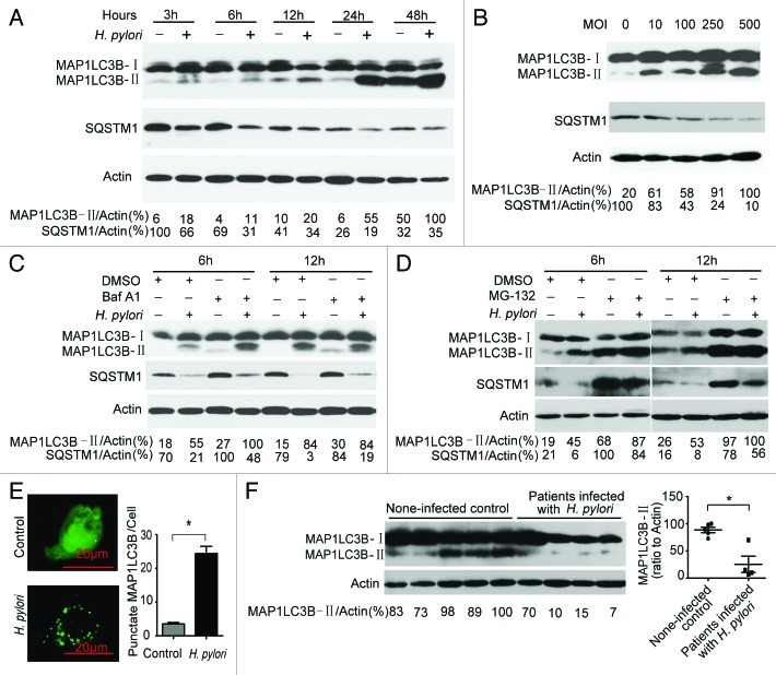
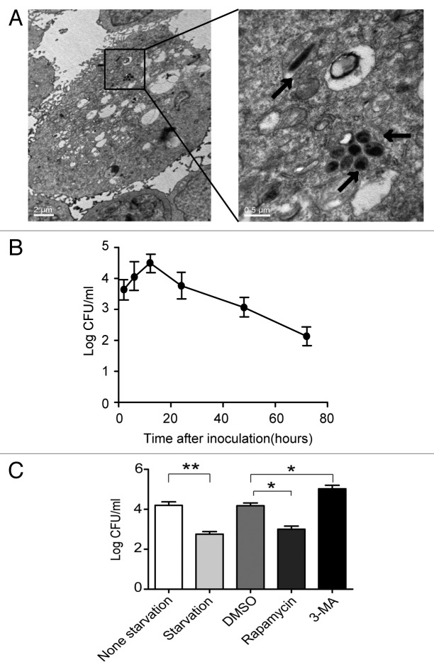
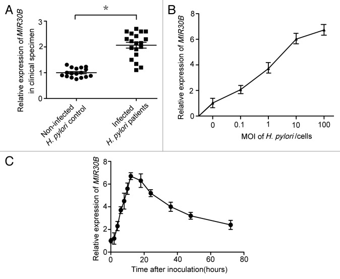
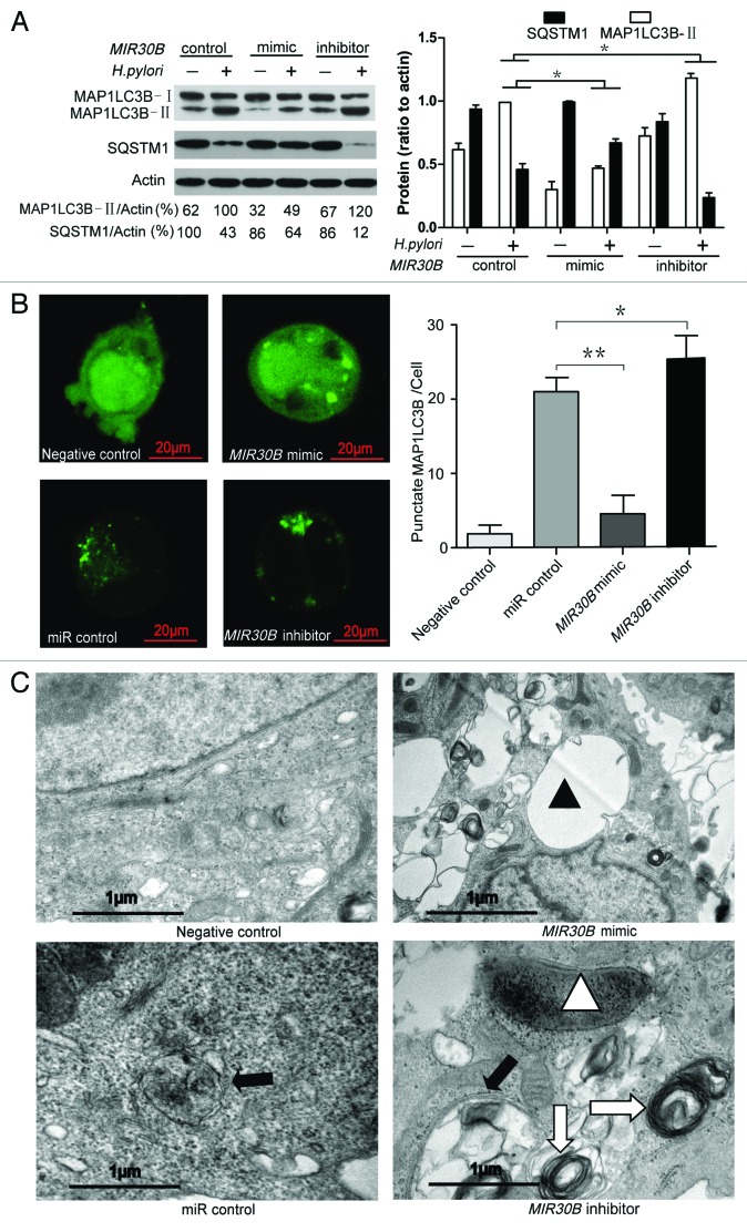
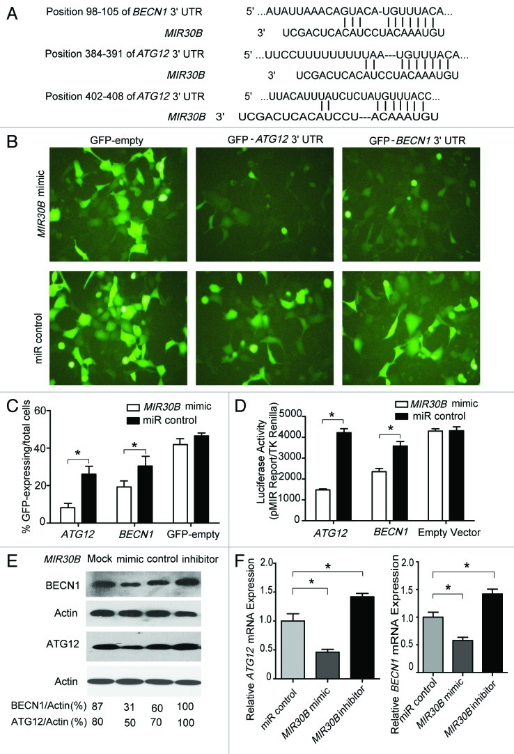
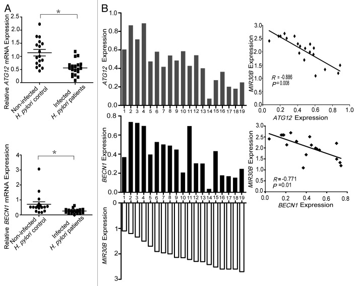
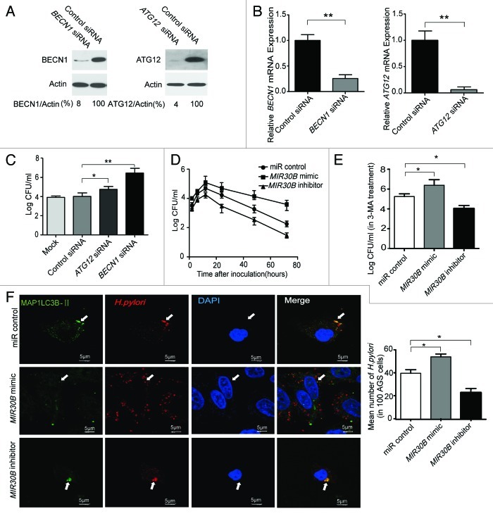
Helicobacter pylori evade immune responses and achieve persistent colonization in the stomach. However, the mechanism by which H. pylori infections persist is not clear. In this study, we showed that MIR30B is upregulated during H. pylori infection of an AGS cell line and human gastric tissues. Upregulation of MIR30B benefited bacterial replication by compromising the process of autophagy during the H. pylori infection. As a potential mechanistic explanation for this observation, we demonstrate that MIR30B directly targets ATG12 and BECN1, which are important proteins involved in autophagy. These results suggest that compromise of autophagy by MIR30B allows intracellular H. pylori to evade autophagic clearance, thereby contributing to the persistence of H. pylori infections.

Keywords: MIR30B; ATG12; BECN1; Helicobacter pylori; autophagy.


Figures

None
Figure 1. Autophagy is induced after H. pylori infection, but it was decreased in patients with chronic H. pylori infection. (A and B) H. pylori increased the conversion of MAP1LC3B-I to MAP1LC3B-II in AGS cells. AGS cells were treated with H. pylori (MOI = 100:1) for 3, 6, 12, 24 and 48 h and treated with H. pylori at MOI = 0, 10, 100, 250 and 500 for 6 h. (C) H. pylori induced incomplete autophagic flux in AGS cells. AGS cells were treated with H. pylori (MOI = 100:1) for 6 h in the presence of Baf A1 (10 nM). (D) AGS cells were treated with H. pylori (MOI = 100:1) for 6 h in the presence of MG-132 (10 μM). (E) AGS cells were transfected with a plasmid expressing GFP-MAP1LC3B. After 24 h, the cells were incubated for 6 h at 37°C in F12 medium with H. pylori. Following fixation, cells were immediately visualized by confocal microscopy. The number of GFP-MAP1LC3B puncta in each cell was counted. (F) The conversion of MAP1LC3B-I to MAP1LC3B-II in gastric mucosal tissues from H. pylori-positive patients (n = 4) was lower than its expression in H. pylori-negative individuals (n = 5). Experiments performed in triplicate showed consistent results. Compared with controls, *p < 0.05.
None
Figure 2. Autophagy inhibition increases multiplication of H. pylori in AGS cells. (A) Representative TEM images of AGS cells after 6 h infection with H. pylori 26695 (MOI = 100:1). Arrows indicate digested bacteria. (B) Multiplication of H. pylori in AGS cells. AGS cells infected with H. pylori (MOI = 100:1) for 3, 6, 12, 24, 48 and 72 h were lysed, and intracellular bacteria were quantified after inoculation. The recovered viable H. pylori were determined as CFU on a CDC anaerobic blood agar plate. (C) The effects of starvation, 3-MA or rapamycin on the multiplication of H. pylori in AGS cells. After pretreatment by starvation, DMSO, rapamycin (100 nM), or 3-MA (2 mM), AGS cells were infected with H. pylori 26695 (MOI = 100:1) for 6 h and then lysed. Intracellular bacteria were quantified after inoculation. *p < 0.05 by Student’s t-test. Results shown are representative of five independent experiments.
None
Figure 3. MIR30B is upregulated in response to H. pylori infection. (A) Expression of MIR30B in gastric mucosal tissues from H. pylori-positive patients (n = 19) was higher than its expression in H. pylori-negative, healthy individuals (n = 17). (B) Real-time PCR detection of MIR30B at indicated MOI after infection of AGS cell lines with H. pylori for 12 h. (C) The kinetics of MIR30B induction assayed by qRT-PCR at different time points (0, 2, 4, 6, 8, 10, 12, 18, 24, 36, 48 and 72 h) after H. pylori stimulation of AGS cells (MOI = 100). Data are representative of five independent experiments. *p < 0.05 via Student’s t-test.
None
Figure 4. MIR30B downregulates autophagy in H. pylori infection. (A) Measurement of MAP1LC3B-II conversion and SQSTM1 degradation with overexpression of MIR30B by western blot assay. AGS cells were transfected with MIR30B control (100 nM), mimic (100 nM) or inhibitor (100 nM) for 24 h and infected with H. pylori for an additional 24 h. Cell lysates were prepared and analyzed. (B) Quantification of GFP-MAP1LC3B puncta (autophagosomes). AGS cells were co-transfected with GFP-MAP1LC3B plasmid and MIR30B control, mimic or inhibitor for 24 h, and were then infected with H. pylori for an additional 6 h. The number of punctate GFP-MAP1LC3B spots in each cell was counted. (C) Ultrastructural alterations in H. pylori-infected AGS cells after transfection with MIR30B control, mimic or inhibitor for 24 h. Infected cells were collected for TEM examination. Negative control shows the mock samples without H. pylori infection. Closed triangles indicate VacA-dependent large vacuoles whereas open triangles show digested bacteria. Closed arrow indicates autophagosomes whereas open arrow shows the multilayer vesicular structure. The asterisks denote significant differences from control (*p < 0.05, **p < 0.01). Experiments performed in triplicate showed consistent results.
None
Figure 5. ATG12 and BECN1 are novel targets of MIR30B. (A) The region of the human ATG12 and BECN1 mRNA 3′UTR predicted to be targeted by MIR30B (TargetScan 4.2). (B and C) Representative fluorescent microscopic image and flow cytometry results demonstrate that GFP expression of the GFP-ATG12 and BECN1 reporters was inhibited by MIR30B. HEK-293 cells were co-transfected with the GFP reporter vectors and compared with cells transfected with a mimic or control of MIR30B. (D) Luciferase reporter assay. HEK293 cells were transiently co-transfected with luciferase reporter vectors and either MIR30B mimic or control. Luciferase activity was normalized to the activity of Renilla luciferase. (E and F) AGS cells were transiently transfected with MIR30B mimic, inhibitor or control for 24 h. The mRNA and protein levels of BECN1 and ATG12 were determined by qRT-PCR and western blot. Data are representative of five independent experiments (* p < 0.05).
None
Figure 6. Inverse correlation between MIR30B and BECN1/ATG12 expression in H. pylori-positive human samples. (A) BECN1/ATG12 and MIR30B expression levels are inversely correlated in human samples. BECN1 and ATG12 mRNA expression in gastric mucosal tissues from H. pylori-positive patients (n = 19) were lower than those of H. pylori-negative individuals (n = 17). (*p < 0.05) (B) The inverse correlation between BECN1/ATG12 and MIR30B expression levels was examined by Spearman correlation analysis (BECN1/MIR30B, R = -0.771, p = 0.01; ATG12/MIR30B, R = -0.886, p = 0.008).
None
Figure 7. MIR30B increases the intracellular survival ratio of H. pylori. (A and B) Detection of inhibition efficiency of siRNAs against BECN1 and ATG12. AGS cells were transfected with two siRNAs targeting BECN1 and ATG12 (100 nM) for 24 h, and mRNA and protein levels of the two genes were determined by qRT-PCR and western blot. (C) Gentamicin protection assay of the intracellular survival ratio of H. pylori with siRNAs against BECN1 and ATG12. AGS cells were transfected with BECN1, ATG12 or a control siRNA at 100 nM for 24 h followed by infection with H. pylori. (D) AGS cells were transfected with MIR30B control, mimic or inhibitor at 100 nM for 24 h, and were then infected with H. pylori for different periods of time (0, 2, 4, 6, 8, 10, 12, 18, 24, 36, 48 and 72 h). Intracellular bacterial counts were determined by gentamicin protection assay. (E) After AGS cells were transfected with MIR30B control, mimic or inhibitor, and pretreated by 3-MA for 24 h, intracellular survival of H. pylori was detected by gentamicin protection assay. (F) H. pylori infection in AGS cells induces GFP-MAP1LC3B puncta formation around the bacteria. AGS cells were transfected by MIR30B control, mimic or inhibitor for 24 h, followed by H. pylori infection for 6 h. The extracellular bacteria were killed with gentamicin. The infected AGS cells were stained with anti-H. pylori antibodies (red) and DAPI (blue). The arrow indicates the presence of H. pylori cells. Data are representative of five independent experiments (*p < 0.05).

References

    1. Suerbaum S, Michetti P. Helicobacter pylori infection. N Engl J Med. 2002;347:1175–86. doi: 10.1056/NEJMra020542. - DOI - PubMed
    1. Acheson DW, Luccioli S. Microbial-gut interactions in health and disease. Mucosal immune responses. Best Pract Res Clin Gastroenterol. 2004;18:387–404. doi: 10.1016/j.bpg.2003.11.002. - DOI - PubMed
    1. Petersen AM, Krogfelt KA. Helicobacter pylori: an invading microorganism? A review. FEMS Immunol Med Microbiol. 2003;36:117–26. doi: 10.1016/S0928-8244(03)00020-8. - DOI - PubMed
    1. Wang YH, Wu JJ, Lei HY. When Helicobacter pylori invades and replicates in the cells. Autophagy. 2009;5:540–2. doi: 10.4161/auto.5.4.8167. - DOI - PubMed
    1. Terebiznik MR, Vazquez CL, Torbicki K, Banks D, Wang T, Hong W, et al. Helicobacter pylori VacA toxin promotes bacterial intracellular survival in gastric epithelial cells. Infect Immun. 2006;74:6599–614. doi: 10.1128/IAI.01085-06. - DOI - PMC - PubMed

Publication types

MeSH terms