. 2012 May 30:9:112.
doi: 10.1186/1742-2094-9-112.

CAR/FoxP3-engineered T regulatory cells target the CNS and suppress EAE upon intranasal delivery

Affiliations

CAR/FoxP3-engineered T regulatory cells target the CNS and suppress EAE upon intranasal delivery

Moa Fransson et al. J Neuroinflammation. .

Abstract

Background: Multiple sclerosis (MS) is an autoimmune disease of the central nervous system (CNS). In the murine experimental autoimmune encephalomyelitis (EAE) model of MS, T regulatory (Treg) cell therapy has proved to be beneficial, but generation of stable CNS-targeting Tregs needs further development. Here, we propose gene engineering to achieve CNS-targeting Tregs from naïve CD4 cells and demonstrate their efficacy in the EAE model.

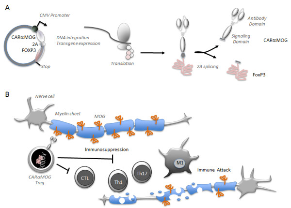
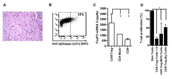
Methods: CD4+ T cells were modified utilizing a lentiviral vector system to express a chimeric antigen receptor (CAR) targeting myelin oligodendrocyte glycoprotein (MOG) in trans with the murine FoxP3 gene that drives Treg differentiation. The cells were evaluated in vitro for suppressive capacity and in C57BL/6 mice to treat EAE. Cells were administered by intranasal (i.n.) cell delivery.

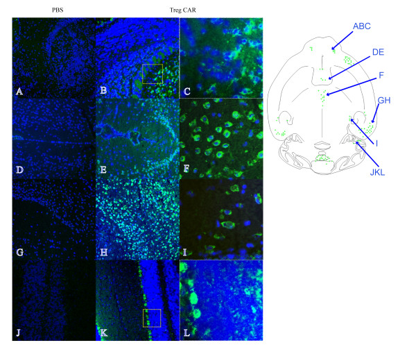
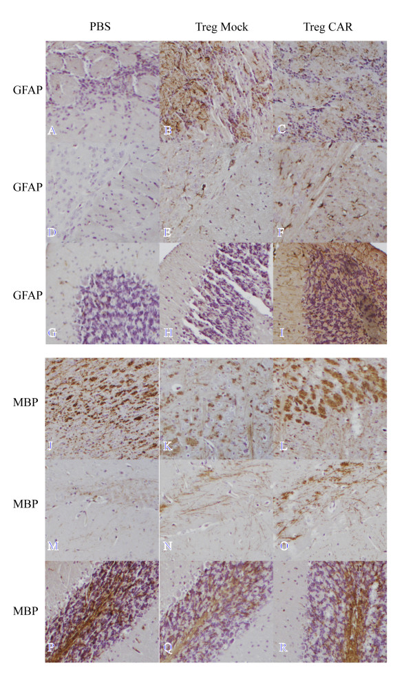
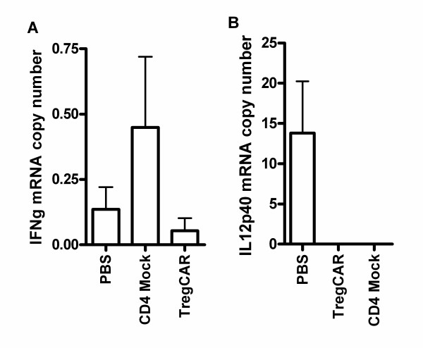
Results: The engineered Tregs demonstrated suppressive capacity in vitro and could efficiently access various regions in the brain via i.n cell delivery. Clinical score 3 EAE mice were treated and the engineered Tregs suppressed ongoing encephalomyelitis as demonstrated by reduced disease symptoms as well as decreased IL-12 and IFNgamma mRNAs in brain tissue. Immunohistochemical markers for myelination (MBP) and reactive astrogliosis (GFAP) confirmed recovery in mice treated with engineered Tregs compared to controls. Symptom-free mice were rechallenged with a second EAE-inducing inoculum but remained healthy, demonstrating the sustained effect of engineered Tregs.

Conclusion: CNS-targeting Tregs delivered i.n. localized to the CNS and efficiently suppressed ongoing inflammation leading to diminished disease symptoms.


Figures

Figure 1
Figure 1
CNS-targeting receptor and FoxP3 translation and function. (A) The CARαMOG-FoxP3 vector contains a scFv cloned from the 8.18 C5 hybridoma. The scFv is linked via an antibody hinge region to the transmembrane and intracellular part of a CD3 zeta chain. The zeta chain is further fused to an intracellular CD28 domain. The murine FoxP3 gene was inserted into the construct after a 2A peptide sequence. Upon translation, the whole expression cassette is translated into a CARFoxP3 fusion protein that is self-cleaved at the 2A site to produce the two separate proteins CARαMOG and FoxP3. (B) CARαMOG and FoxP3 is transported to the cell surface and nucleus, respectively. At the cell surface CARαMOG can bind to MOG + cells to attach the Treg to those cells and prevent immune attacks on MOG + cells such as oligodendrocytes in the CNS. FoxP3 will drive the Treg phenotype by regulating gene transcription in the nucleus.
Figure 2
Figure 2
Engineered Tregs express CAR and FoxP3. (A) Antibodies from the 8.18 C5 hybridoma cross-reacted with murine myelin as demonstrated by immunohistochemistry (red colour expression). (B) CD4+T cells transduced with CARαMOG-FoxP3 lentivectors (CAR Tregs) were stained with fluorescein isothiocyanate (FITC)-conjugated antibodies and analyzed for surface expression of the scFv by flow cytometry. (C) FoxP3 copy number in transduced cells analyzed by quantitative PCR analysis. Expression of FoxP3 is significantly higher than both CD4-mock and naïve CD4+T cells (P < 0,05) as analyzed by Mann-Whitney using GraphPad prism software. The expression experiments were repeated for every cell generation with similar result. (D) CAR Tregs were mixed with αCD3/IL-2-stimulated T cells at a1:2 ratio and analyzed for suppressive ability in a thymidine-based assay. CAR Tregs were able to suppress activated T-cells (P < 0.05). CAR Tregs suppressed αCD3/IL-2-stimulated T cells in the presence of activated macrophages (P < 0.05) or MOG-expressing cells (P < 0.05). The experiments were repeated and statistical differences analyzed by Mann-Whitney test using GraphPad prism software.
Figure 3
Figure 3
Engineered Tregs localize to the CNS when administered intranasally in naive mice. Treg cells were administered intranasally in the right nostril and the distribution of green fluorescent protein (GFP) in horizontal cryosections of the brain of naïve mice was studied 24 hours after the delivery. The schematic drawing describes a selective immunofluorescence in various brain regions (green spots). GFP immunofluorescence is present in the granular and to a lower extent in the external plexiform layer of the olfactory bulb (B, C), lateral septal nucleus (E), central medial thalamic nucleus (F), ectorhinal cortex (H), medial genic nucleus (I) and cerebellum (K, L) of a CAR Treg-treated naïve mouse. Corresponding areas showing no GFP fluorescence in a PBS-treated naïve mouse are (A, D, G, J). Enlargements of areas in olfactory bulb and cerebellum (as indicated by boxes) are depicted in C and L. Detail of GFP immunofluorescence in the central medial thalamic nucleus and medial genic nucleus (F, I). Cell nuclei (blue) are stained with DAPI Original magnification 10× (A, B, D, E, G, H, J, K) and 40× (C, F, I, L).
Figure 4
Figure 4
CNS-targeting Tregs can reduce EAE symptoms. (A) Ten mice in three groups were given 1 × 105 CAR Tregs, CD4+ Mock T cells or PBS alone by i.n. administration at the peak of EAE inflammation (15 days post-EAE immunization) and thereafter were monitored for EAE symptoms. Ten days post cell treatment all EAE mice in the CAR Treg group were cured (P < 0.001). At end point (15 days post cell treatment) four out of ten EAE mice in the Mock-treated group still exhibited EAE symptoms. The experiment was repeated three times with similar results. (B) Symptom-free mice from each treatment group were given a second dose of EAE-inducing inoculum and monitored for EAE symptoms. CAR Treg-treated mice were able to resist EAE inflammation to a higher extent than CD4+ Mock-treated mice (P < 0.001). Pooled data from six EAE mice (three/group from two separate experiments) are shown in the figure. Scores for individual mice are shown separately in the figure. (C) Ten EAE mice in three groups were administered 1x10⁵ CAR Tregs, CD4+ Mock T cells or PBS alone by i.p. injection at peak of EAE inflammation (day 15). At end point (day 15 post-treatment) all mice in the CAR Τreg group were cured but six out of ten mice in the Mock CD4+ T cell group still exhibited EAE symptoms. Statistics are analyzed with Mann-Whitney test using GraphPad prism software. *P < 0.05, **P < 0.01, ***P < 0.001.
Figure 5
Figure 5
Astrogliosis and remyelination after intranasal administration of CNS-targeting Treg. Mice in three groups were given 1 × 105 CAR Tregs, CD4+ Mock T cells or PBS alone by intranasal administration at the peak of EAE inflammation (15 days post-EAE immunization). Fifteen days post cell treatment the mice were killed and brain sections from each group (CAR Tregs, Mock CD4+ T cell and PBS) were analyzed for reactive astroglisosis using glial acidic fibrillary protein (GFAP) (A-I) and myelination using myelin basic protein (MBP) (J-R). GFAP was evaluated in olfactory bulb (A-C), corpus callosum (D-F), and cerebellum (G-I) of brain sagittal section from PBS, Mock CD4+ T cell and CAR Τreg-treated EAE mice. In the olfactory bulb of mice treated with Mock CD4+ T cells there was a strong staining for GFAP whereas this area in PBS-treated mice (A) and of CAR Treg-treated mice (C) exhibited a weak and moderate staining, respectively. There is an extremely weak staining in PBS-treated EAE mice, moderate staining in Mock CD4+ T cell-treated EAE mice and strong staining in CAR Treg-treated EAE mice for corpus callosum and cerebellum. MBP was evaluated in brain stem (J-L), hippocampus (M-O) and cerebellum (P-R) of brain sagittal sections in PBS, Mock CD4+ T cell and CAR Treg-treated EAE mice. In the brain stem and cerebellum of mice treated with CAR Tregs, there was a moderate staining for MBP (L, R) whereas the brain stem of Mock-treated mice (K,Q) and PBS-treated mice (J,P) exhibited a weak and strong staining, respectively. There is an extremely weak staining in PBS-treated EAE mice, a moderate staining in Mock CD4+ T cell-treated EAE mice and strong staining in CAR Treg-treated EAE mice for hippocampus. Original magnification 10×.
Figure 6
Figure 6
Decreased expression of effector cytokines in CNS-targeting CAR Treg-treated brain. Mice in three groups were given 1 × 105 CAR Tregs, CD4+ Mock T cells or PBS alone by i.n. administration at the peak of EAE inflammation (15 days post-EAE immunization). Fifteen days post cell treatment, brain biopsies from five EAE mice per group (CAR Tregs, Mock CD4+ T cells and PBS) were analyzed for expression of effector cytokines (IL-12 and IFNγ) by quantitative RT-PCR. Error bars represent standard error of the mean (SEM).

References

    1. Martin R, McFarland HF. Immunological aspects of experimental allergic encephalomyelitis and multiple sclerosis. Crit Rev Clin Lab Sci. 1995;32:121–182. doi: 10.3109/10408369509084683. - DOI - PubMed
    1. Maloy KJ, Powrie F. Regulatory T cells in the control of immune pathology. Nat Immunol. 2001;2:816–822. doi: 10.1038/ni0901-816. - DOI - PubMed
    1. Coutinho A, Hori S, Carvalho T, Caramalho I, Demengeot J. Regulatory T cells: the physiology of autoreactivity in dominant tolerance and “quality control” of immune responses. Immunol Rev. 2001;182:89–98. doi: 10.1034/j.1600-065X.2001.1820107.x. - DOI - PubMed
    1. Ziegler SF. FOXP3: of mice and men. Annu Rev Immunol. 2006;24:209–226. doi: 10.1146/annurev.immunol.24.021605.090547. - DOI - PubMed
    1. Sakaguchi S, Sakaguchi N. Regulatory T cells in immunologic self-tolerance and autoimmune disease. Int Rev Immunol. 2005;24:211–226. doi: 10.1080/08830180590934976. - DOI - PubMed

Publication types

MeSH terms

Substances