Longitudinal progression of frontal and temporal lobe changes in schizophrenia
- PMID: 22647883
- PMCID: PMC3413315
- DOI: 10.1016/j.schres.2012.05.002
Longitudinal progression of frontal and temporal lobe changes in schizophrenia
Abstract
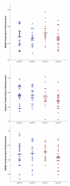
Cortical abnormalities are considered a neurobiological characteristic of schizophrenia. However, the pattern of such deficits as they progress over the illness remains poorly understood. The goal of this project was to assess the progression of cortical thinning in frontal and temporal cortical regions in schizophrenia, and determine whether relationships exist between them and neuropsychological and clinical symptom profiles. As part of a larger longitudinal 2-year follow-up study, schizophrenia (n=20) and healthy participants (n=20) group-matched for age, gender, and recent-alcohol use, were selected. Using MRI, estimates of gray matter thickness were derived from primary anatomical gyri of the frontal and temporal lobes using surface-based algorithms. These values were entered into repeated-measures analysis of variance models to determine group status and time effects. Change values in cortical regions were correlated with changes in neuropsychological functioning and clinical symptomatology. Results revealed exaggerated cortical thinning of the middle frontal, superior temporal, and middle temporal gyri in schizophrenia participants. These thickness changes strongly influenced volumetric reductions, but were not related to shrinking surface area. Neuropsychological and clinical symptom profiles were stable in the schizophrenia participants despite these neuroanatomic changes. Overall it appears that ongoing abnormalities in the cerebral cortex continue after initial onset of schizophrenia, particularly the lateral aspects of frontal and temporal regions, and do not relate to neuropsychological or clinical measures over time. Maintenance of neuropsychological performance and clinical stability in the face of changing neuroanatomical structure suggests the involvement of alternative compensatory mechanisms.
Copyright © 2012 Elsevier B.V. All rights reserved.
Figures
Similar articles
-
Frontal and temporal lobe brain volumes in schizophrenia. Relationship to symptoms and clinical subtype.Arch Gen Psychiatry. 1995 Dec;52(12):1061-70. doi: 10.1001/archpsyc.1995.03950240079013. Arch Gen Psychiatry. 1995. PMID: 7492258
-
Changes in the frontotemporal cortex and cognitive correlates in first-episode psychosis.Biol Psychiatry. 2010 Jul 1;68(1):51-60. doi: 10.1016/j.biopsych.2010.03.019. Epub 2010 May 10. Biol Psychiatry. 2010. PMID: 20452574 Free PMC article.
-
Dynamically spreading frontal and cingulate deficits mapped in adolescents with schizophrenia.Arch Gen Psychiatry. 2006 Jan;63(1):25-34. doi: 10.1001/archpsyc.63.1.25. Arch Gen Psychiatry. 2006. PMID: 16389194
-
[Hypofrontality and negative symptoms in schizophrenia: synthesis of anatomic and neuropsychological knowledge and ecological perspectives].Encephale. 2001 Sep-Oct;27(5):405-15. Encephale. 2001. PMID: 11760690 Review. French.
-
Familial and developmental abnormalities of front lobe function and neurochemistry in schizophrenia.J Psychopharmacol. 1997;11(2):133-42. doi: 10.1177/026988119701100206. J Psychopharmacol. 1997. PMID: 9254279 Review.
Cited by
-
Early-course unmedicated schizophrenia patients exhibit elevated prefrontal connectivity associated with longitudinal change.J Neurosci. 2015 Jan 7;35(1):267-86. doi: 10.1523/JNEUROSCI.2310-14.2015. J Neurosci. 2015. PMID: 25568120 Free PMC article.
-
Patterns of regional gray matter loss at different stages of schizophrenia: A multisite, cross-sectional VBM study in first-episode and chronic illness.Neuroimage Clin. 2016 Jun 3;12:1-15. doi: 10.1016/j.nicl.2016.06.002. eCollection 2016. Neuroimage Clin. 2016. PMID: 27354958 Free PMC article.
-
Brain transcriptomic profiling reveals common alterations across neurodegenerative and psychiatric disorders.Comput Struct Biotechnol J. 2022 Aug 19;20:4549-4561. doi: 10.1016/j.csbj.2022.08.037. eCollection 2022. Comput Struct Biotechnol J. 2022. PMID: 36090817 Free PMC article.
-
Cortical connectomic mediations on gamma band synchronization in schizophrenia.Transl Psychiatry. 2023 Jan 19;13(1):13. doi: 10.1038/s41398-022-02300-6. Transl Psychiatry. 2023. PMID: 36653335 Free PMC article.
-
A Systematic Review of Cognition-Brain Morphology Relationships on the Schizophrenia-Bipolar Disorder Spectrum.Schizophr Bull. 2021 Oct 21;47(6):1557-1600. doi: 10.1093/schbul/sbab054. Schizophr Bull. 2021. PMID: 34097043 Free PMC article.
References
-
- Albus M, Hubmann W, Scherer J, Dreikorn B, Hecht S, Sobizack N, et al. A prospective 2-year follow-up study of neurocognitive functioning in patients with first-episode schizophrenia. Eur Arch Psychiatry Clin Neurosci. 2002;252(6):262–267. - PubMed
-
- Andreasen NC. The Scale for the Assessment of Negative Symptoms (SANS) The University of Iowa; Iowa City, IA: 1983.
-
- Andreasen NC. The Scale for the Assessment of Positive Symptoms (SAPS) The University of Iowa; Iowa City, IA: 1984.
-
- Antonova E, Sharma T, Morris R, Kumari V. The relationship between brain structure and neurocognition in schizophrenia: A selective review. Schizophr Res. 2004;70(2-3):117–145. - PubMed
Publication types
MeSH terms
Substances
Grants and funding
LinkOut - more resources
Full Text Sources
Medical
Research Materials


