. 2012 Jul;60(1):154-62.
doi: 10.1161/HYPERTENSIONAHA.111.189688. Epub 2012 May 29.

Activation of Nod-like receptor protein 3 inflammasomes turns on podocyte injury and glomerular sclerosis in hyperhomocysteinemia

Affiliations

Activation of Nod-like receptor protein 3 inflammasomes turns on podocyte injury and glomerular sclerosis in hyperhomocysteinemia

Chun Zhang et al. Hypertension. 2012 Jul.

Abstract

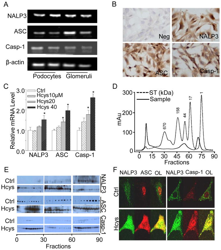
Inflammasome is a multiprotein complex consisting of Nod-like receptor protein 3 (NALP3), apoptosis-associated speck-like protein (ASC), and caspase 1 or 5, which functions to switch on the inflammatory process. The present study hypothesized that the formation and activation of NALP3 inflammasomes turn on podocyte injury leading to glomerulosclerosis during hyperhomocysteinemia (hHcys). RT-PCR and Western blot analysis demonstrated that murine podocytes expressed 3 essential components of the NALP3 inflammasome complex, namely, NALP3, ASC, and caspase 1. Treatment of podocytes with l-homocysteine induced the formation of NALP3 inflammasome complex, an increase in caspase 1 activity, podocyte cytoskeleton rearrangement, and decreased production of vascular endothelial growth factor from podocytes, which were all blocked by silencing the ASC gene or inhibiting caspase 1 activity. In mice with hHcys induced by feeding them a folate-free diet, NALP3 inflammasome formation and activation in glomerular podocytes were detected at an early stage, as shown by confocal microscopy, size exclusion chromatography of the assembled inflammasome complex, and increased interleukin-1β production in glomeruli. Locally silencing the ASC gene in the kidney significantly reduced NALP3 inflammasome formation and interleukin 1β production in glomeruli of mice with hHcys. Pathologically, hHcys-associated albuminuria, foot process effacement of podocytes, loss of podocyte slit diaphragm molecules, and glomerulosclerosis at the late stage were significantly improved by local ASC gene silencing or by caspase 1 inhibition. In conclusion, NALP3 inflammasome formation and activation on stimulation of homocysteine are important molecular mechanisms triggering podocyte injury and ultimately resulting in glomerulosclerosis in hHcys.


Figures

Figure 1
Figure 1. Hcys-induced activation of NALP3 inflammasomes in cultured podocytes
A. RT-PCR gel document showing the expression of NALP3, ASC, and caspase-1 in cultured podocytes. B. Immunocytochemical staining of NALP3, ASC, and caspase-1 in podocytes (original magnification, ×100). C. mRNA expression of NALP3, ASC, and caspase-1 in podocytes with or without stimulation of L-Hcys (n=6). D. Elution profile of proteins (optical density at 280 nm) from podocytes by SEC. The molecular mass scale was determined using a wide range of molecular mass standard. ST=standard. E. SDS-PAGE of proteins fractions via SEC from control and Hcys-treated podocytes that were probed with anti-NALP3, ASC, or casapse-1 antibodies, respectively. F. Representative confocal microscopic images showing colocalization of NALP3 (green) with ASC (red) or NALP3 (green) with caspase-1 (red, Casp-1) in podocytes (original magnification, ×400). OL=overlay. * P<0.05 vs. control.
Figure 2
Figure 2. Effects of ASC gene silencing and caspase-1 inhibition on inflammasome activation and functional changes in podocytes
A. Representative confocal microscopic images showing colocalization of NALP3 (green) with ASC (red) or caspase-1 (red, Casp-1) in podocytes (original magnification, ×400), OL=overlay. B. Caspase-1 activity. C. IL-1β production. D. VEGF-A secretion in podocytes with or without treatment of L-Hcys and/or ASC shRNA and caspase-1 inhibitor. Ctrl: control; Veh: vehicle; Scra: scrambled siRNA; Casp-1: Caspase-1; WEHD: Z-WEHD-FMK, n=5. * P<0.05 vs. control; # P<0.05 vs. Hcys.
Figure 3
Figure 3. Formation and activation of podocyte NALP3 inflammasomes during hHcys in mice
A. Colocalization of NALP3 (green) with ASC (red) or caspase-1 (red) in mouse glomeruli on the normal diet or folate free diet (FF diet). Casp-1: Caspase-1. B. Summarized data showing the fold changes in PCC for the colocalization of NALP3 with ASC or caspase-1 (n=6). C. Colocalization of podocin (red) with NALP3 (green) or caspase-1 (green) in mouse glomeruli. D. Summarized data showing the fold changes in PCC for the colocalization of podocin with NALP3 or caspase-1 (n=6). E. Caspase-1 activity, F. IL-1β concentrations in mouse glomeruli (n=6) * P<0.05 vs. 0 week.
Figure 4
Figure 4. Effects of ASC shRNA or caspase-1 inhibition on podocyte inflammasome formation and activation during hHcys
A. Colocalization of NALP3 (green) with ASC (red) or caspase-1 (red) in mouse glomeruli. B. Summarized data showing the fold changes in PCC for the colocalization of NALP3 with ASC or caspase-1 (n=6). C. Caspase-1 activity in different groups of mice (n=6). D. IL-1β production in mouse glomeruli (n=6 per group). N Diet: Normal Diet; Casp-1: Caspase-1; Scra: Scrambled shRNA-transfected; ASC sh: ASC shRNA-transfected. *P<0.05, vs. scrambled shRNA-transfected mice on the N diet; # P<0.05, vs. scrambled shRNA-transfected mice on the FF diet.
Figure 5
Figure 5. Effects of ASC gene silencing and caspase-1 inhibition on hHcys-induced glomerular damage
A. Urinary albumin excretion in 6 groups of mice as indicated (n=8 per group). B. PAS staining showing glomerular morphological changes (original magnification, ×400). C. Summarized data of glomerular damage index in 6 different groups of mice (n=6 per group). D. ASC gene silencing and caspase-1 inhibition improved podocyte ultrastructure in FF diet-treated mice as shown by TEM examination. Arrow denotes the area of foot process effacement in WT mice on the FF diet (n=3). Original magnification: ×8,000.

References

    1. Mariathasan S, Newton K, Monack DM, Vucic D, French DM, Lee WP, Roose-Girma M, Erickson S, Dixit VM. Differential activation of the inflammasome by caspase-1 adaptors asc and ipaf. Nature. 2004;430:213–218. - PubMed
    1. Zhou R, Tardivel A, Thorens B, Choi I, Tschopp J. Thioredoxin-interacting protein links oxidative stress to inflammasome activation. Nat Immunol. 2010;11:136–140. - PubMed
    1. Imaeda AB, Watanabe A, Sohail MA, Mahmood S, Mohamadnejad M, Sutterwala FS, Flavell RA, Mehal WZ. Acetaminophen-induced hepatotoxicity in mice is dependent on tlr9 and the nalp3 inflammasome. J Clin Invest. 2009;119:305–314. - PMC - PubMed
    1. Iyer SS, Pulskens WP, Sadler JJ, Butter LM, Teske GJ, Ulland TK, Eisenbarth SC, Florquin S, Flavell RA, Leemans JC, Sutterwala FS. Necrotic cells trigger a sterile inflammatory response through the nlrp3 inflammasome. Proc Natl Acad Sci U S A. 2009;106:20388–20393. - PMC - PubMed
    1. Niemir ZI, Stein H, Dworacki G, Mundel P, Koehl N, Koch B, Autschbach F, Andrassy K, Ritz E, Waldherr R, Otto HF. Podocytes are the major source of il-1 alpha and il-1 beta in human glomerulonephritides. Kidney Int. 1997;52:393–403. - PubMed

Publication types

MeSH terms