

Review
doi: 10.1097/RMR.0b013e31825c062c.
Molecular characterization of rheumatoid arthritis with magnetic resonance imaging
Affiliations
- PMID: 22648081
- PMCID: PMC3387734
- DOI: 10.1097/RMR.0b013e31825c062c
Item in Clipboard
Review
Molecular characterization of rheumatoid arthritis with magnetic resonance imaging
Top Magn Reson Imaging.
2011 Apr.
Abstract
Several recent advances in the field of magnetic resonance imaging (MRI) may transform the detection and monitoring of rheumatoid arthritis (RA). These advances depict both anatomic and molecular alterations from RA. Previous techniques could detect specific end products of metabolism in vitro or were limited to providing anatomic information. This review focuses on the novel molecular imaging techniques of hyperpolarized carbon-13 MRI, MRI with iron-labeled probes, and fusion of MRI with positron emission tomography. These new imaging approaches go beyond the anatomic description of RA and lend new information into the status of this disease by giving molecular information.
Figures
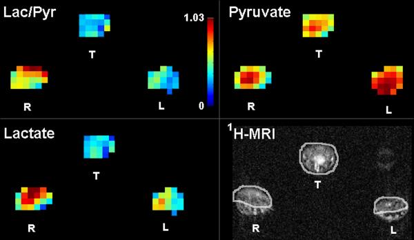
Quantitative metabolic maps in an arthritic rat after injection of hyperpolarized 13C-pyruvate show increased lactate production in the arthritic right paw as measured by the lactate-to-pyruvate ratio (Lac/Pyr). The color bar indicates relative levels of Lac/Pyr. Red color is the maximum signal intensity for 13C-pyruvate = 1485 au, 13C-lactate = 674 au, and Lac/Pyr = 1.03. 1H-MRI shows soft tissue swelling in the arthritic right (R) paw in comparison to the control left (L) paw and the region of interest analysis applied for the metabolic maps. T=tail, au=arbitrary units. Reproduced with permission from MacKenzie JD, et al. Radiology 2011.

Spectroscopic profiles from single 2.5 × 2.5 × 10 mm voxels in control and arthritic tissues in one rat. Increased 13C-lactate production is demonstrated in the plantar surface of the arthritic right paw (lactate-to-pyruvate ratio = 0.70) in comparison to the control left paw (= 0.57), tail (= 0.37), and tissues away from the site of arthritis induction in the dorsal right paw (= 0.57). Arrow indicates alanine production in the control left paw, but no alanine is observed in the arthritic paw. Reproduced with permission from MacKenzie JD, et al. Radiology 2011.
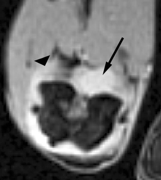
Transverse T2*-weighted fast gradient-echo images (1000/15; flip angle, 25°) show T2* effects at USPIO-enhanced MR imaging of macrophage activity in antigen-induced arthritis in rabbits. (a) Precontrast image shows joint effusion (arrow) that is surrounded by a thickened synovium (arrowhead). (b) Postcontrast image (24 hours after USPIO administration) shows susceptibility effects (arrow) within the synovium (arrowhead), representing USPIO uptake in phagocytic-active macrophages. Signal intensity of bone marrow (*) is decreased in comparison to that in a, which is caused by USPIO uptake by the mononuclear phagocyte system within the bone marrow. Reproduced with permission from Lutz AM, et al. Radiology 2004.
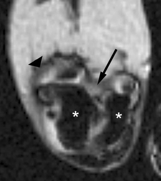
Transverse T2*-weighted fast gradient-echo images (1000/15; flip angle, 25°) show T2* effects at USPIO-enhanced MR imaging of macrophage activity in antigen-induced arthritis in rabbits. (a) Precontrast image shows joint effusion (arrow) that is surrounded by a thickened synovium (arrowhead). (b) Postcontrast image (24 hours after USPIO administration) shows susceptibility effects (arrow) within the synovium (arrowhead), representing USPIO uptake in phagocytic-active macrophages. Signal intensity of bone marrow (*) is decreased in comparison to that in a, which is caused by USPIO uptake by the mononuclear phagocyte system within the bone marrow. Reproduced with permission from Lutz AM, et al. Radiology 2004.

Fusion of high-resolution 18F-FDG-PET with MRI for an RA therapy responder. Images were generated using the clinical MRI and high resolution PET/CT sequential scanning approach. The top row shows images from baseline while the bottom row depicts images at 4 weeks after the initiation of treatment. Figures (a) and (c) depict representative axial sections from a pre-contrast T1-weighted MR image of the patient's wrist, (b) and (d) show the same section from the T1-weighted MR image acquired post-administration of a gadolinium-based contrast agent, and (c) and (f) demonstrate the MRI-PET fusion images of the same section. Red arrows show synovitis in the carpal region, green arrows indicate inflammation at sites of erosions while the blue arrows indicate inflammation at the base of the thumb (corresponding to osteoarthritis). Dramatic reductions in synovitis and metabolic activity at sites of erosions were measured from MRI-PET at 4 weeks and correlated with the rheumatologist's score obtained at the patient's 3-month standard-of-care visit.
Similar articles
-
Screening for peptides targeted to IL-7Rα for molecular imaging of rheumatoid arthritis synovium.Arthritis Res Ther. 2016 Oct 12;18(1):230. doi: 10.1186/s13075-016-1133-8. Arthritis Res Ther. 2016. PMID: 27729062 Free PMC article.
-
Detection of subclinical synovitis with macrophage targeting and positron emission tomography in patients with rheumatoid arthritis without clinical arthritis.J Rheumatol. 2014 Nov;41(11):2145-52. doi: 10.3899/jrheum.140059. Epub 2014 Oct 1. J Rheumatol. 2014. PMID: 25274888
-
Macrophages mediated diagnosis of rheumatoid arthritis using fibrin based magnetic nanoparticles as MRI contrast agents.Biochim Biophys Acta Gen Subj. 2017 Jan;1861(1 Pt A):2992-3001. doi: 10.1016/j.bbagen.2016.09.018. Epub 2016 Sep 20. Biochim Biophys Acta Gen Subj. 2017. PMID: 27663233
-
Emerging optical and nuclear medicine imaging methods in rheumatoid arthritis.Nat Rev Rheumatol. 2012 Dec;8(12):719-28. doi: 10.1038/nrrheum.2012.148. Epub 2012 Sep 25. Nat Rev Rheumatol. 2012. PMID: 23007740 Review.
-
Ultrasound versus high field magnetic resonance imaging in rheumatoid arthritis.Clin Exp Rheumatol. 2014 Jan-Feb;32(1 Suppl 80):S99-105. Epub 2014 Feb 17. Clin Exp Rheumatol. 2014. PMID: 24528508 Review.
Cited by
-
Focusing on ligamentous soft tissue inflammation for the future understanding of early axial psoriatic arthritis.Rheumatology (Oxford). 2024 Dec 1;63(Supplement_2):ii7-ii14. doi: 10.1093/rheumatology/keae568. Rheumatology (Oxford). 2024. PMID: 39700474 Free PMC article. Review.
-
Advancing musculoskeletal research with nanoscience.Nat Rev Rheumatol. 2013 Oct;9(10):614-23. doi: 10.1038/nrrheum.2013.112. Epub 2013 Jul 23. Nat Rev Rheumatol. 2013. PMID: 23881069 Review.
-
In vivo quantification of mouse autoimmune arthritis by PET/CT.Int J Rheum Dis. 2016 May;19(5):452-8. doi: 10.1111/1756-185X.12410. Epub 2014 Jun 26. Int J Rheum Dis. 2016. PMID: 24965561 Free PMC article.
-
Hyperpolarized 13 C magnetic resonance imaging for noninvasive assessment of tissue inflammation.NMR Biomed. 2021 Mar;34(3):e4460. doi: 10.1002/nbm.4460. Epub 2020 Dec 8. NMR Biomed. 2021. PMID: 33291188 Free PMC article. Review.
-
EXPLORing Arthritis with Total-body Positron Emission Tomography.Semin Musculoskelet Radiol. 2023 Dec;27(6):632-640. doi: 10.1055/s-0043-1775746. Epub 2023 Nov 7. Semin Musculoskelet Radiol. 2023. PMID: 37935209 Free PMC article. Review.
References
-
- MacKenzie JD, Karasick D. Imaging of Rheumatoid Arthritis. In: Weissman B, editor. Imaging of Arthritis and Metabolic Bone Disease. Elsevier Health Sciences; Philadelphia: 2009. pp. 340–64.
-
- Sommer OJ, Kladosek A, Weiler V, Czembirek H, Boeck M, Stiskal M. Rheumatoid Arthritis: A Practical Guide to State-of-the-Art Imaging, Image Interpretation, and Clinical Implications1. Radiographics. 2005;25:381–98. - PubMed
-
- McQueen FM, Ostergaard M. Established rheumatoid arthritis - new imaging modalities. Best Pract Res Clin Rheumatol. 2007;21:841–56. - PubMed
-
- Scott DL, Kingsley GH. Tumor necrosis factor inhibitors for rheumatoid arthritis. N Engl J Med. 2006;355:704–12. - PubMed
-
- Felson DT, Anderson JJ, Boers M, et al. American College of Rheumatology. Preliminary definition of improvement in rheumatoid arthritis. Arthritis Rheum. 1995;38:727–35. - PubMed
Publication types
MeSH terms
Substances
Grants and funding
LinkOut - more resources
Full Text Sources
Medical
