. 2012 Jul;23(14):2712-22.
doi: 10.1091/mbc.E12-03-0228. Epub 2012 May 30.

Efficient secretion of small proteins in mammalian cells relies on Sec62-dependent posttranslational translocation

Affiliations

Efficient secretion of small proteins in mammalian cells relies on Sec62-dependent posttranslational translocation

Asvin K K Lakkaraju et al. Mol Biol Cell. 2012 Jul.

Abstract

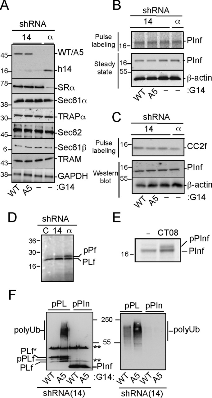
Mammalian cells secrete a large number of small proteins, but their mode of translocation into the endoplasmic reticulum is not fully understood. Cotranslational translocation was expected to be inefficient due to the small time window for signal sequence recognition by the signal recognition particle (SRP). Impairing the SRP pathway and reducing cellular levels of the translocon component Sec62 by RNA interference, we found an alternate, Sec62-dependent translocation path in mammalian cells required for the efficient translocation of small proteins with N-terminal signal sequences. The Sec62-dependent translocation occurs posttranslationally via the Sec61 translocon and requires ATP. We classified preproteins into three groups: 1) those that comprise ≤100 amino acids are strongly dependent on Sec62 for efficient translocation; 2) those in the size range of 120-160 amino acids use the SRP pathway, albeit inefficiently, and therefore rely on Sec62 for efficient translocation; and 3) those larger than 160 amino acids depend on the SRP pathway to preserve a transient translocation competence independent of Sec62. Thus, unlike in yeast, the Sec62-dependent translocation pathway in mammalian cells serves mainly as a fail-safe mechanism to ensure efficient secretion of small proteins and provides cells with an opportunity to regulate secretion of small proteins independent of the SRP pathway.


Figures

FIGURE 1:
FIGURE 1:
Cells with an impaired SRP pathway translocate preproinsulin and pre-CC chemokine 2 efficiently. (A) Cellular levels of different SRP and membrane components after shRNA(14) and shRNA(SRα) expression in HeLa cells. Cell extracts were obtained at 72 h posttransfection. (B, C) Translocation of the reporter proteins indicated was assessed by pulse-labeling experiments (top) and by Western blotting of total cell extracts after treatment with MG132 (10 μm) for 8 h (bottom). In pulse-labeling experiments, cells were treated with 10 μM MG132 during depletion of cold amino acids and during labeling. (B, C) pInf and pCC2f in h14k.d. or SRαk.d. HeLa cells. Wild type and A5: h14k.d. cells expressing the fusion proteins G14 and G14A5, respectively. (D) Pulse labeling of preprolactin in h14k.d. and SRαk.d. HeLa cells as well as control cells expressing shRNA(luc). (E) pInf translocation in wild-type HeLa cells and cells treated with the Sec61 translocon inhibitor CT08. (F) Polyubiquitination of preprolactin and preproinsulin in h14k.d. cells and complemented as in B. Cells also expressed HA-tagged ubiquitin. Immunoprecipitation of the reporter proteins was done with anti-FLAG antibodies. Left and right, Western blots using both anti-FLAG and anti-HA or only anti-HA antibodies, respectively. PLf*, phosphorylated forms of prolactin in transit for secretion; **light and heavy chains of the antibodies.
FIGURE 2:
FIGURE 2:
Characterization of Sec62 knockdown in HeLa cells. (A) Western blots of extracts from HeLa cells expressing shRNA 62.2 (left) and shRNAs 62.3 and 14 (right). Membranes were probed with antibodies against the proteins indicated. Glyceraldehyde 3-phosphate dehydrogenase (GAPDH) and β-actin, loading controls. 62.2 and 62.3, two different shRNAs targeting Sec62 mRNA. Arrow, time point of all following experiments. (B) HeLa cells expressing shRNAs (62.3) and the negative control (Luc) were analyzed for cell growth by counting the number of live cells manually every 24 h starting from 48 h posttransfection. Error bars, SD. (C) G protein expression after infection of Sec62k.d. and control HeLa cells with vesicular stomatitis virus. The cells were permeabilized with Triton to reveal the G protein in the secretory pathway. Scale bar, 10 μm. (D) Western blots of cell extracts from cells expressing shRNA 62.2 (left) and shRNAs Sec62.3 and 14 (right) probed with antibodies against the proteins indicated. Cal, calnexin. The quantified values varied between 90 and 100% in single-knockdown and between 86 and 115% in double-knockdown experiments when normalized to GAPDH and compared with control cells.
FIGURE 3:
FIGURE 3:
SRP-independent translocation depends on Sec62. (A) Protein translocation of preproinsulin (pPInf), pre-CC2 (pCC2f), and preprolactin (pPLf) monitored in cells depleted of either of h14 and Sec62 individually (lanes 2 and 3) or of h14 and Sec62 simultaneously (lanes 4–7) after treatment with MG132 (10 μM) for 8 h. Lane 1, shRNA(Luc). The double-knockdown cells were complemented by the expression of G14 (lane 5), G14A5 (lane 6), and Sec62 (lane 7). Reporter proteins were revealed with anti-FLAG antibody. PLf*, phosphorylated prolactin in transit for secretion. (B) Total cell extracts from the experiment shown in A revealed by Western blotting for the proteins indicated. (C) Polyubiquitination of nontranslocated pInf. Western blots of total cell extracts prepared from HeLa cells expressing shRNAs 62.3 and 14 and, when indicated, G14 and G14A5 after treatment with MG132 for 8 h. Cells also expressed HA-tagged ubiquitin. Insulin was immunoprecipitated and revealed with anti-FLAG, polyubiquitination of insulin was revealed with anti-HA, and fusion proteins in cell extracts were revealed with anti-GFP antibodies.
FIGURE 4:
FIGURE 4:
Sec62-dependent translocation is efficient for preproteins comprising <160 amino acids. Translocation efficiencies (T[%]) of different reporter proteins in HeLa cells with reduced cellular levels of Sec62 (A) and SRα (B). C, shRNA(Luc). Pulse-labeled proteins were immunoprecipitated with anti-FLAG antibodies, displayed by SDS–PAGE, and visualized by phosphoimager. Nontranslocated preproteins were stabilized with MG132 (10 μM) before and during labeling. Proteins are named according to their chain length (see Table 1). p, preprotein. (C) SRP-dependent translocation of preprolactin proteins of decreasing chain length in HEK 293T cells. Cells expressed shRNA (14) and, as indicated, G14 and G14A5. Preprotein accumulation was monitored by Western blotting of cell extracts harvested after treatment with MG132 (10 μM) for 8 h. To resolve the signal sequence–cleaved and uncleaved forms, the proteins were displayed on different gels. pPLf and pPLf-194, 12%; pPLf-163 and pPLf-140, 15%. (D) Preresistin accumulation in Sec62k.d. cells. Western blots of cell extracts with anti-His antibodies. Cells were treated with the protease inhibitor ALLN (50 μM) for 8 h before harvesting. (E) Polyubiquitination of preprolactin proteins of different chain length in HeLa cells expressed shRNA 14 and, as indicated, G14 and G14A5. Cells also expressed HA-tagged ubiquitin. Left, total cell extracts probed with anti-FLAG antibodies to reveal the reporter proteins. Right, reporter proteins were immunoprecipitated with anti-FLAG antibodies and displayed by SDS–PAGE, and the reporter proteins and ubiquitination were revealed by Western blotting with anti-FLAG and anti-HA antibodies, respectively.
FIGURE 5:
FIGURE 5:
Posttranslational translocation in vitro requires Sec62. In vitro synthesized 35S-labeled preproteins (Input) were incubated with permeabilized HeLa cells, cytosolic extract, and ATP for translocation. (A) pPIn (left) and pPLf-190 and pPLf-140 (right) in the presence or absence of ATP. Translocation was monitored by signal sequence cleavage (lane 3). (B) pPIn translocation with and without CT08. (C) pPIn, pCC2 incubated with permeabilized SRα and control HeLa cells. (D) pPIn translocation with cytosol from h14k.d. cells. (E) pPIn, pLCh, and pRes translocation with permeabilized Sec62k.d. (+) and control (–) HeLa cells. The relative translocation efficiencies (percentages) as compared with the inputs are shown in parentheses.
FIGURE 6:
FIGURE 6:
Sec62 can be immunoprecipitated with a short reporter protein. HeLa cells were transfected with pRh-100– and pPLf-expressing plasmids, cells were harvested 25 h later after treatment with cycloheximide (3 μg/ml) for 1 h, and protein complexes were precipitated with the antibodies indicated. F, anti-FLAG antibody; H, anti-His antibody. (A) Immunoprecipitates displayed on a 4–20% gradient gel. The upper part of the membrane was revealed with anti-Sec62 and the lower part with anti-His antibodies. pRh-100 and Rh-100 could not be resolved on this gel. Right, total cell extracts displayed on Tris-Tricine gel and 12% PAGE for Rh-100 and Sec62, respectively. (B) As in A, but with preprolactin as a reporter protein. Protein was revealed with anti-FLAG antibody. PL*, phosphorylated form of prolactin; **heavy chain.

References

    1. Brodsky JL, Goeckeler J, Schekman R. BiP and Sec63p are required for both co- and posttranslational protein translocation into the yeast endoplasmic reticulum. Proc Natl Acad Sci USA. 1995;92:9643–9646. - PMC - PubMed
    1. Chen Y, Zhang Y, Yin Y, Gao G, Li S, Jiang Y, Gu X, Luo J. SPD—a Web-based secreted protein database. Nucleic Acids Res. 2005;33:D169–D173. - PMC - PubMed
    1. Cross BC, Sinning I, Luirink J, High S. Delivering proteins for export from the cytosol. Nat Rev Mol Cell Biol. 2009;10:255–264. - PubMed
    1. Daimon M, Susa S, Suzuki K, Kato T, Yamatani K, Sasaki H. Identification of a human cDNA homologue to the Drosophila translocation protein 1 (Dtrp1) Biochem Biophys Res Commun. 1997;230:100–104. - PubMed
    1. Deshaies RJ, Sanders SL, Feldheim DA, Schekman R. Assembly of yeast Sec proteins involved in translocation into the endoplasmic reticulum into a membrane-bound multisubunit complex. Nature. 1991;349:806–808. - PubMed

Publication types