. 2012 Aug;23(15):2905-16.
doi: 10.1091/mbc.E11-12-1007. Epub 2012 May 30.

The clathrin adaptor Dab2 recruits EH domain scaffold proteins to regulate integrin β1 endocytosis

Affiliations

The clathrin adaptor Dab2 recruits EH domain scaffold proteins to regulate integrin β1 endocytosis

Anjali Teckchandani et al. Mol Biol Cell. 2012 Aug.

Abstract

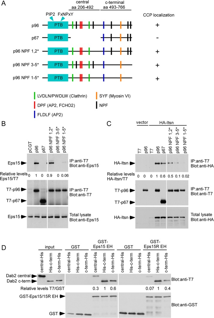
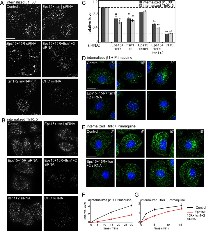
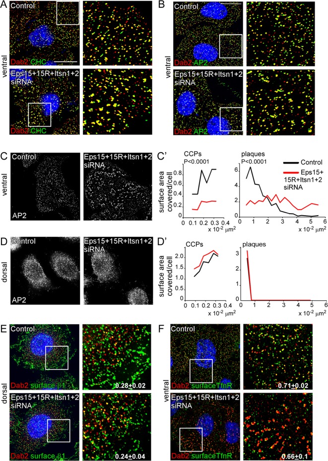
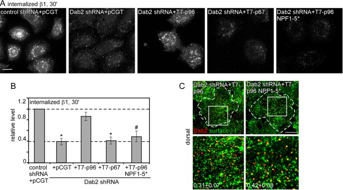
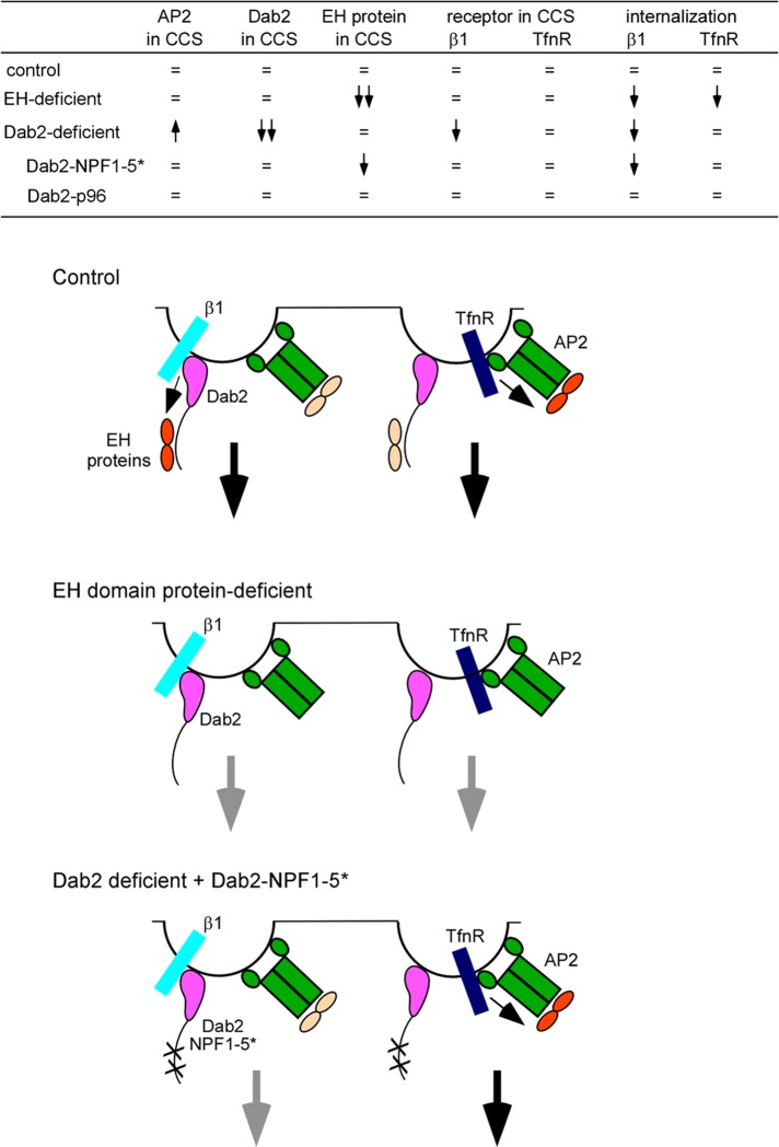
Endocytic adaptor proteins facilitate cargo recruitment and clathrin-coated pit nucleation. The prototypical clathrin adaptor AP2 mediates cargo recruitment, maturation, and scission of the pit by binding cargo, clathrin, and accessory proteins, including the Eps-homology (EH) domain proteins Eps15 and intersectin. However, clathrin-mediated endocytosis of some cargoes proceeds efficiently in AP2-depleted cells. We found that Dab2, another endocytic adaptor, also binds to Eps15 and intersectin. Depletion of EH domain proteins altered the number and size of clathrin structures and impaired the endocytosis of the Dab2- and AP2-dependent cargoes, integrin β1 and transferrin receptor, respectively. To test the importance of Dab2 binding to EH domain proteins for endocytosis, we mutated the EH domain-binding sites. This mutant localized to clathrin structures with integrin β1, AP2, and reduced amounts of Eps15. Of interest, although integrin β1 endocytosis was impaired, transferrin receptor internalization was unaffected. Surprisingly, whereas clathrin structures contain both Dab2 and AP2, integrin β1 and transferrin localize in separate pits. These data suggest that Dab2-mediated recruitment of EH domain proteins selectively drives the internalization of the Dab2 cargo, integrin β1. We propose that adaptors may need to be bound to their cargo to regulate EH domain proteins and internalize efficiently.


Figures

FIGURE 1:
FIGURE 1:
The Dab2 NPFs are required for binding Eps15 and Itsn. (A) Drawing of T7-p96, T7-p67, or T7-p96 NPF mutants showing binding sites. The Dab2 central and C-terminal fragments used for in vitro binding are also shown. (B, C) HEK 293 cells were transfected with control vector or T7-p96, T7-p67, or T7-p96 NPF mutants and HA-Itsn. Cell lysates were immunoprecipitated with anti-T7 antibody and immunoblotted with either anti-Eps15 (B) or anti-HA (C). Representative blots from one of four independent experiments are shown. Pixel intensities were measured by ImageJ. (D) Binding of purified Dab2 and Eps15/15R EH domain. Purified, bacterially grown T7-mDab2-206-492-His (central fragment), T7-mDab2-493-766-His (C-terminus of Dab2), and T7-His-mDab2-493-766 (C-terminus of Dab2 with N-terminal tag) were mixed with purified, glutathione-Sepharose–bound GST-EH domain of Eps15 or Eps15R. Pixel intensities were measured by ImageJ.
FIGURE 2:
FIGURE 2:
EH domain proteins regulate integrin β1 and TfnR endocytosis. (A–C) Control and siRNA-treated HeLa cells were incubated with anti–integrin β1 (A) or anti-TfnR antibody (B) for 30 min at 4°C, followed by warming at 37°C for 30 (β1) or 5 min (TfnR). (D–G) Control and EH domain–deficient HeLa cells were incubated with anti–integrin β1 (D) or anti-TfnR antibody (E) for 30 min at 4°C and then warmed to 37°C for the indicated times in the presence of 2 μM Primaquine. Surface antibody was removed by acid stripping, and internalized antibody was detected. (A, B, D, E) Z-projections of the entire cell. (C, F, G) Pixel intensities measured by ImageJ. Mean values and standard errors of internalized receptor after normalizing with surface receptor at time = 0 are shown. Approximately 15 cells/treatment from three independent experiments were analyzed. #p < 0.05, *p < 0.01, **p < 0.001 by t test. Bar, 10 μm.
FIGURE 3:
FIGURE 3:
Effect of EH domain protein depletion on clathrin, AP2, Dab2, and receptor localization. Control and EH domain–deficient HeLa cells grown on collagen IV–coated coverslips were fixed, permeabilized, and stained with anti-Dab2 and anti-CHC (A) or anti-AP2 (B) to detect clathrin-coated structures. (C) AP2 staining alone on the ventral and dorsal surface. AP2 staining was used to calculate the surface area covered by CCPs (size, 0.009–0.032 μm2, which corresponds to structures with diameters 107–202 nm) or plaques (size, >0.032 μm2) by ImageJ. Approximately 2000 puncta/treatment from three independent experiments were used. p values were calculated using the Mann–Whitney test, a nonparametric test. (E, F) Cells were stained with either anti–integrin β1 (E) or anti-TfnR (F) antibody before permeabilizing to detect surface receptor. Shown are 0.2-μm sections of the ventral or dorsal surface. The white boxes indicate the enlarged images shown in the insets. The fraction of surface integrin β1 or TfnR that colocalized with Dab2 was measured by ImageJ (Manders colocalization coefficient). In all cases random colocalization, obtained by flipping the red channel (Dab2), was <0.08. Approximately 10 cells/treatment from three separate experiments were analyzed. Bar, 10 μm.
FIGURE 4:
FIGURE 4:
Disruption of the Dab2–EH domain protein interaction slows Dab2-dependent integrin β1 internalization. (A–C) Control and Dab2-deficient HeLa cells reexpressing vector alone or T7-p96, T7-p67, or T7-p96 NPF 1-5* were incubated with anti–integrin β1 antibody for 30 min at 4°C, followed by warming at 37°C for 30 min. (A, B) Surface antibody was removed by acid stripping, and internalized antibody was detected. (A) Z-projections of the entire cell showing internalized integrin β1 antibody. (B) Pixel intensities were measured by ImageJ. Mean values and standard errors are shown for ∼30 cells/treatment from three independent experiments. #p < 0.05, *p < 0.01, by t test. (C) Cells were stained with anti–integrin β1 antibody before permeabilizing to detect surface receptor. Z-projections of the dorsal surface are shown. The white boxes indicate the enlarged images shown in the insets. Bar, 10 μm. The fraction of surface integrin β1 that colocalized with Dab2 in T7-p96 or T7-p96 NPF 1-5*–expressing cells was calculated using ImageJ (Manders colocalization coefficient). In all cases random colocalization, obtained by flipping the red channel (Dab2), was <0.07. Approximately five cells/treatment were analyzed.
FIGURE 5:
FIGURE 5:
Disruption of the Dab2-EH domain protein interaction does not affect TfnR endocytosis. (A, B) Control and Dab2-deficient HeLa cells reexpressing vector alone or T7-p96, T7-p67, or T7-p96 NPF 1-5* were incubated with anti-TfnR antibody for 30 min at 4°C, followed by warming at 37°C for 5 min. (A, B) Surface antibody was removed by acid stripping, and internalized antibody was detected. (A) Z-projections of the entire cell showing internalized TfnR antibody. (B) Pixel intensities were measured by ImageJ. Mean values and standard errors are shown for ∼30 cells/treatment from three independent experiments. #p < 0.05, *p < 0.01, by t test. (C) Control cells were incubated with anti–integrin β1 antibody and Alexa Fluor 488–conjugated human Tfn to detect surface β1 and TfnR or permeabilized and stained with anti-Dab2 and anti-AP2. Z-projections of the dorsal surface are shown. The white boxes indicate the enlarged images shown in the insets. Bar, 10 μm. The fraction of CCSs that contain both receptors integrin β1 and Tfn or both adaptors Dab2 and AP2 is shown. Random colocalization, obtained by flipping the red channel, was <0.03.
FIGURE 6:
FIGURE 6:
Summary of results and proposed model. Top, summary of results taken from Table 2, Supplemental Figure S2, and Figures 2, 4, and 5. =, same as in control; upward arrow, increased; downward arrow, decreased. Bottom, model. Dab2 and AP2 are found together in most clathrin-coated structures. However, their receptors, integrin β1 and TfnR, do not localize to the same structures. Internalization of a clathrin structure is driven by adaptors bound to both receptor and EH domain proteins (shown in orange). Inactive EH domains are shown in pale yellow. Small black arrows indicate regulation of EH domain proteins by adaptor–receptor complexes. Large black arrows indicate endocytosis. In the absence of EH domain proteins internalization of integrin β1 and TfnR is impaired (gray arrows). When cells are reconstituted with a Dab2 mutant that does not bind EH domain proteins, integrin β1 is not internalized efficiently because this Dab2 mutant cannot bind EH domain proteins. However, TfnR endocytosis proceeds normally because its adaptor AP2 can bind EH domain proteins. The AP2–EH domain complex in integrin β1 structures is unable to drive endocytosis probably because it is not bound to receptor.

Similar articles

Cited by

References

    1. Bleil JD, Bretscher MS. Transferrin receptor and its recycling in HeLa cells. EMBO J. 1982;1:351–355. - PMC - PubMed
    1. Boettner DR, D'Agostino JL, Torres OT, Daugherty-Clarke K, Uygur A, Reider A, Wendland B, Lemmon SK, Goode BL. The F-BAR protein Syp1 negatively regulates WASp-Arp2/3 complex activity during endocytic patch formation. Curr Biol. 2009;19:1979–1987. - PMC - PubMed
    1. Cao H, Chen J, Krueger EW, McNiven MA. SRC-mediated phosphorylation of dynamin and cortactin regulates the “constitutive” endocytosis of transferrin. Mol Cell Biol. 2010;30:781–792. - PMC - PubMed
    1. Cao TT, Mays RW, von Zastrow M. Regulated endocytosis of G-protein-coupled receptors by a biochemically and functionally distinct subpopulation of clathrin-coated pits. J Biol Chem. 1998;273:24592–24602. - PubMed
    1. Chao WT, Kunz J. Focal adhesion disassembly requires clathrin-dependent endocytosis of integrins. FEBS Lett. 2009;583:1337–1343. - PMC - PubMed

Publication types

MeSH terms