. 2013 Mar;9(1):25-33.
doi: 10.1007/s13181-012-0239-x.

The effect of curcumin on oxaliplatin and cisplatin neurotoxicity in rats: some behavioral, biochemical, and histopathological studies

Affiliations

The effect of curcumin on oxaliplatin and cisplatin neurotoxicity in rats: some behavioral, biochemical, and histopathological studies

Mansour S Al Moundhri et al. J Med Toxicol. 2013 Mar.

Abstract

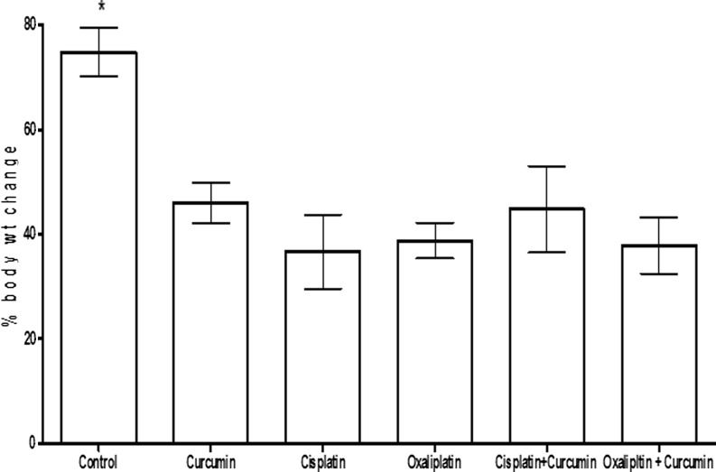
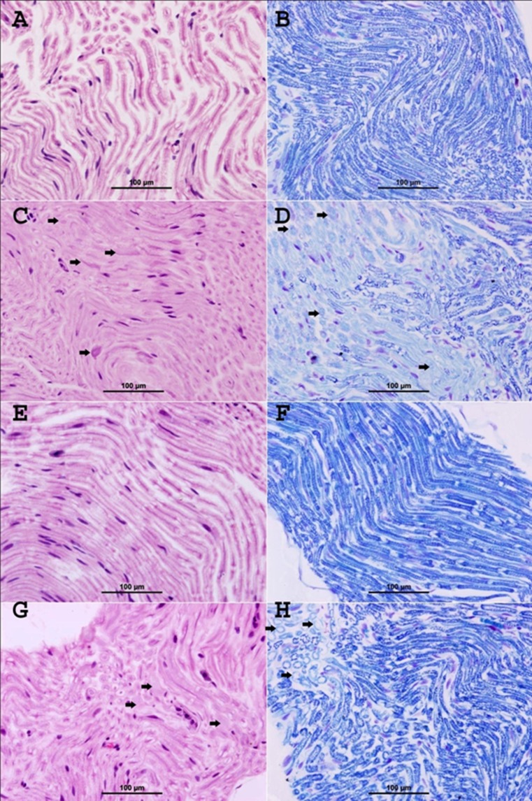
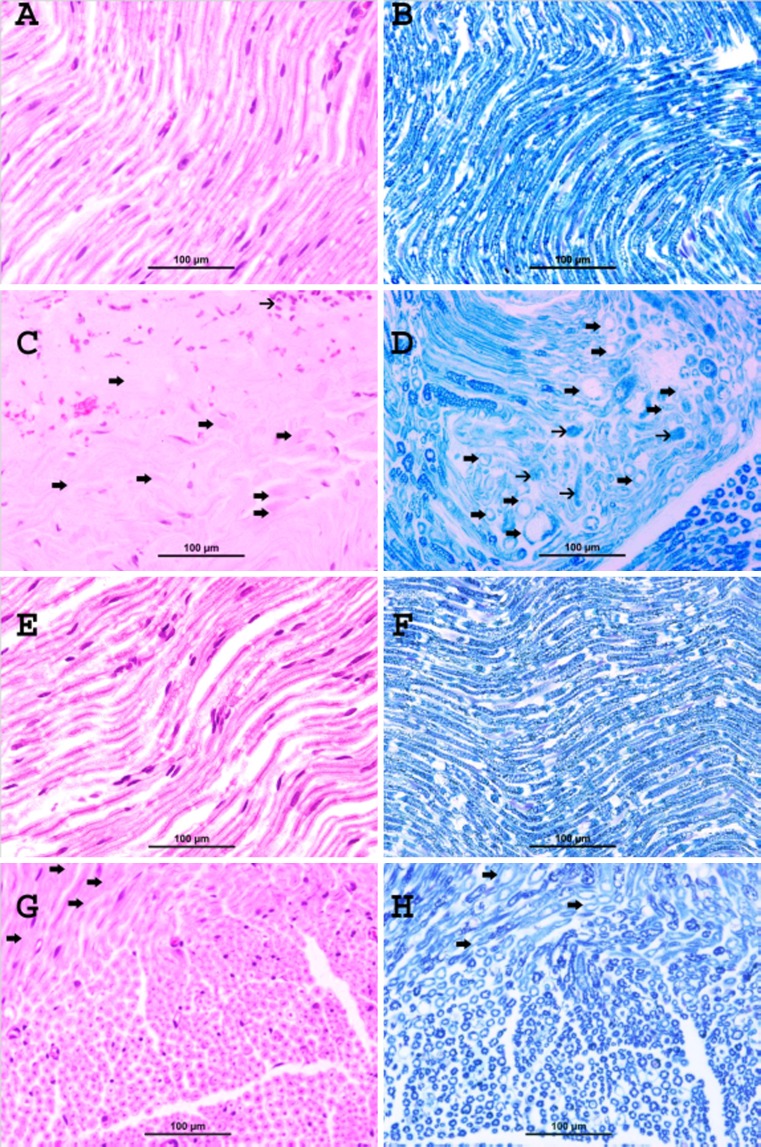
Cisplatin is commonly used against several solid tumors, and oxaliplatin is an effective cytotoxic drug used in colorectal cancer. A major clinical issue affecting 10-40 % of patients treated with cisplatin or oxaliplatin is severe peripheral neuropathy causing sensory, motor, and autonomic dysfunction, with symptoms including cold sensitivity and neuropathic pain. The biochemical basis of the neurotoxicity is uncertain, but is associated with oxidative stress. Curcumin (a natural phenolic yellow pigment) has strong antioxidant, anticancer, and anti-inflammatory actions. Here we report the possible protective effect of curcumin on some cisplatin- and oxaliplatin-induced behavioral, biochemical, and histopathological alterations in rats. Twenty-four hours after the end of treatments some motor and behavioral tests (motor activity, thermal and mechanical nociception, and neuromuscular coordination) were conducted, followed by measuring plasma neurotensin platinum concentration in the sciatic nerve, and studying the histopathology of the sciatic nerve. Oxaliplatin (4 mg/kg) and cisplatin (2 mg/kg) [each given twice weekly, in a total of nine intraperitoneal injections over 4.5 weeks] significantly increased plasma neurotensin concentration, caused specific damage in the histology of the sciatic nerve and produced variable effects in the motor and behavioral tests. Oral curcumin (10 mg/kg, 4 days before the platinum drug, and thereafter, concomitantly with it for 4.5 weeks) reversed the alterations in the plasma neurotensin and sciatic nerve platinum concentrations, and markedly improved sciatic nerve histology in the platinum-treated rats. Larger experiments using a wider dose range of oxaliplatin, cisplatin, and curcumin are required to fully elucidate the possible protective role of curcumin in platinum-induced neurotoxicity.


Figures

Fig. 1
Fig. 1
The percentage body increase over the period of study (4.5 weeks) in rats treated with glucose (1 ml/kg, twice weekly; a total of nine injections in 4.5 weeks), curcumin (10 mg/kg, given orally 4 days prior to the platinum drug, and thereafter, concomitantly with the drug for 4.5 weeks), cisplatin (2 mg/kg, twice weekly; a total of nine injections in 4.5 weeks), oxaliplatin (4 mg/kg, twice weekly; a total of nine injections in 4.5 weeks), and cisplatin + curcumin and oxaliplatin + curcumin, at the above doses. Each column and vertical bar depict mean ± SEM (n = 6 rats). Growth in the control group was significantly higher than in the rest of the groups (among which there was no significant difference in body weight increase)
Fig. 2
Fig. 2
Representative photographs of sections of the sciatic nerve of rats treated with glucose (A and B), cisplatin (C and D), curcumin (E and F), and cisplatin + curcumin (G and H), as follows: A control glucose-treated group shows normal myelinated nerve fibers,(H&E stain). B Control glucose-treated group shows deep blue staining of normal myelin sheath (LFB stain). C Cisplatin-treated group shows focal areas of demyelination and degeneration of the nerve fibers (thick arrows). Areas of infiltration with mononuclear inflammatory cells were also noticed (thin arrows) (H&E stain). D Cisplatin-treated group demonstrates areas of demyelination (thick arrows), which have light blue staining compared with the normal deep blue staining in normal fibers, and decrease in the caliber of nerve fibers. Foci of axonal degeneration were also seen (thin arrows) (LFB stain). E Curcumin-treated group shows normal appearance of myelinated nerve fibers (H&E stain). F Curcumin-treated group shows normal deep blue staining of the myelin sheath (LFB stain). G Cisplatin + curcumin-treated group shows marked decrease in the demyelination (arrows) (H&E stain). H Cisplatin + curcumin-treated group demonstrates a marked decrease of demyelination (arrows), which have light blue staining compared with the normal deep blue staining in normal fibers (LFB stain)
Fig. 3
Fig. 3
Representative photographs of sections of the sciatic nerve of rats treated with glucose (A and B), oxaliplatin (C and D), curcumin (E and F), and oxaliplatin + curcumin (G and H) as follows: A control glucose-treated group shows normal myelinated nerve fibers (H&E stain). B Control glucose-treated group shows deep blue staining of normal myelin sheath (LFB stain). C Oxaliplatin-treated group shows focal areas of demyelination and degeneration of the nerve fibers (thick arrows). Areas of infiltration with mononuclear inflammatory cells were also noticed (thin arrows) (H&E stain). D Oxaliplatin-treated group demonstrates areas of demyelination (thick arrows), which have light blue staining compared with the normal deep blue staining in normal fibers, and decrease in the caliber of nerve fibers. Foci of axonal degeneration were also seen (thin arrows) (LFB stain). E Curcumin-treated group shows normal appearance of myelinated nerve fibers (H&E stain). F Curcumin-treated group shows normal deep blue staining of the myelin sheath (LFB stain). G Oxaliplatin + curcumin-treated group shows marked decrease in the demyelination (arrows) (H&E stain). H Oxaliplatin + curcumin-treated group demonstrates a marked decrease of demyelination (arrows), which have light blue staining compared with the normal deep blue staining in normal fibers (LFB stain)

References

    1. Saif MW, Reardon J. Management of oxaliplatin-induced peripheral neuropathy. Ther Clin Risk Manag. 2005;1:249–58. - PMC - PubMed
    1. Shah N, Dizon DS. New-generation platinum agents for solid tumors. Future Oncol. 2009;5:33–42. doi: 10.2217/14796694.5.1.33. - DOI - PubMed
    1. Grothey A. Clinical management of oxaliplatin-associated neurotoxicity. Clin Colorectal Cancer. 2005;5(Suppl 1):S38–46. doi: 10.3816/CCC.2005.s.006. - DOI - PubMed
    1. Broomand A, Jerremalm E, Yachnin J, Ehrsson H, Elinder F. Oxaliplatin neurotoxicity—no general ion channel surface-charge effect. J Negat Results Biomed. 2009;8:2. doi: 10.1186/1477-5751-8-2. - DOI - PMC - PubMed
    1. Cavaletti G, Marmiroli P. Chemotherapy-induced peripheral neurotoxicity. Nature Review Neurology. 2011;6:657–666. doi: 10.1038/nrneurol.2010.160. - DOI - PubMed

Publication types

MeSH terms