

Trabecular level analysis of bone cement augmentation: a comparative experimental and finite element study
- PMID: 22648574
- PMCID: PMC3438401
- DOI: 10.1007/s10439-012-0587-3
Trabecular level analysis of bone cement augmentation: a comparative experimental and finite element study
Abstract
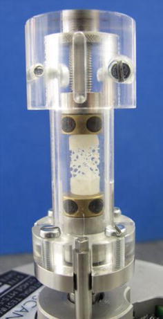
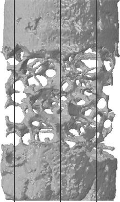
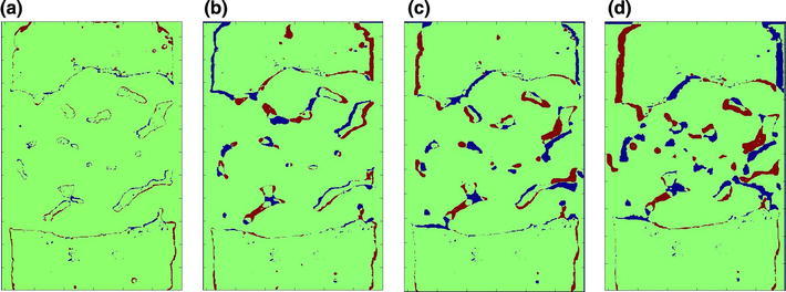
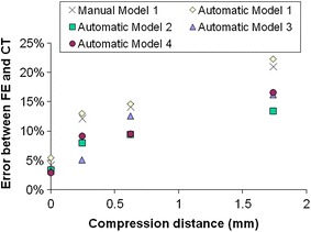
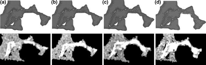
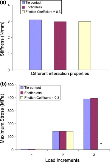
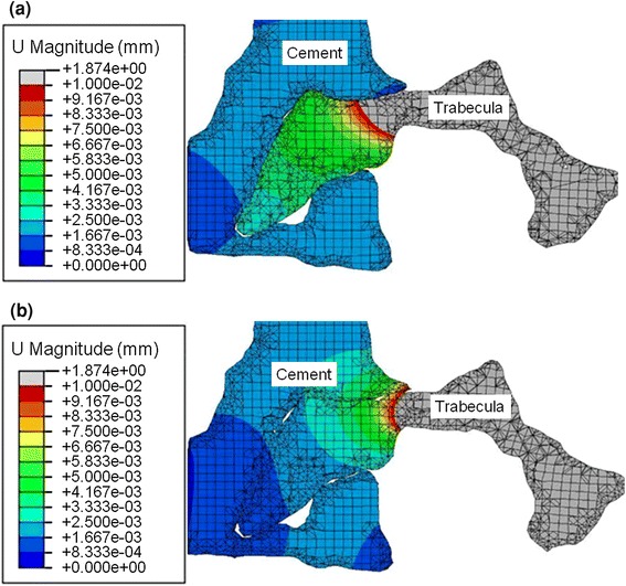
The representation of cement-augmented bone in finite element (FE) models of vertebrae following vertebroplasty remains a challenge, and the methods of the model validation are limited. The aim of this study was to create specimen-specific FE models of cement-augmented synthetic bone at the microscopic level, and to develop a new methodology to validate these models. An open cell polyurethane foam was used reduce drying effects and because of its similar structure to osteoporotic trabecular bone. Cylindrical specimens of the foam were augmented with PMMA cement. Each specimen was loaded to three levels of compression inside a micro-computed tomography (μCT) scanner and imaged both before compression and in each of the loaded states. Micro-FE models were generated from the unloaded μCT images and displacements applied to match measurements taken from the images. A morphological comparison between the FE-predicted trabecular deformations and the corresponding experimental measurements was developed to validate the accuracy of the FE model. The predicted deformation was found to be accurate (less than 12% error) in the elastic region. This method can now be used to evaluate real bone and different types of bone cements for different clinical situations.
Figures
Similar articles
-
Modelling cement augmentation: a comparative experimental and finite element study at the continuum level.Proc Inst Mech Eng H. 2010;224(7):903-11. doi: 10.1243/09544119JEIM696. Proc Inst Mech Eng H. 2010. PMID: 20839657
-
Biomechanical evaluation of calcium phosphate-based nanocomposite versus polymethylmethacrylate cement for percutaneous kyphoplasty.Spine J. 2019 Nov;19(11):1871-1884. doi: 10.1016/j.spinee.2019.06.007. Epub 2019 Jun 14. Spine J. 2019. PMID: 31202837
-
Development of specimen-specific finite element models of human vertebrae for the analysis of vertebroplasty.Proc Inst Mech Eng H. 2008 Feb;222(2):221-8. doi: 10.1243/09544119JEIM285. Proc Inst Mech Eng H. 2008. PMID: 18441757
-
The mechanical behavior of PMMA/bone specimens extracted from augmented vertebrae: a numerical study of interface properties, PMMA shrinkage and trabecular bone damage.J Biomech. 2012 May 11;45(8):1478-84. doi: 10.1016/j.jbiomech.2012.02.012. Epub 2012 Mar 2. J Biomech. 2012. PMID: 22386105
-
Long-term effects of vertebroplasty: adjacent vertebral fractures.J Long Term Eff Med Implants. 2006;16(4):265-80. doi: 10.1615/jlongtermeffmedimplants.v16.i4.10. J Long Term Eff Med Implants. 2006. PMID: 17073569 Review.
Cited by
-
Peri-implant stress correlates with bone and cement morphology: Micro-FE modeling of implanted cadaveric glenoids.J Orthop Res. 2015 Nov;33(11):1671-9. doi: 10.1002/jor.22933. Epub 2015 Jun 18. J Orthop Res. 2015. PMID: 25929691 Free PMC article.
-
Development and validation of a nomogram for predicting new vertebral compression fractures after percutaneous kyphoplasty in postmenopausal patients.J Orthop Surg Res. 2023 Nov 30;18(1):914. doi: 10.1186/s13018-023-04400-5. J Orthop Surg Res. 2023. PMID: 38037128 Free PMC article.
-
Characterisation of a metallic foam-cement composite under selected loading conditions.J Mater Sci Mater Med. 2013 Nov;24(11):2509-18. doi: 10.1007/s10856-013-5000-8. Epub 2013 Jul 12. J Mater Sci Mater Med. 2013. PMID: 23846838
-
Biomechanical evaluation of screw and cement placement strategies for treating medial uncontained tibial defects in total knee arthroplasty: A finite element analysis.Med Int (Lond). 2024 Jun 25;4(5):47. doi: 10.3892/mi.2024.171. eCollection 2024 Sep-Oct. Med Int (Lond). 2024. PMID: 38983796 Free PMC article.
-
Finite element study on the micromechanics of cement-augmented proximal femoral nail anti-rotation (PFNA) for intertrochanteric fracture treatment.Sci Rep. 2024 May 6;14(1):10322. doi: 10.1038/s41598-024-61122-2. Sci Rep. 2024. PMID: 38710745 Free PMC article.
References
Publication types
MeSH terms
Substances
Grants and funding
LinkOut - more resources
Full Text Sources
Medical
