. 2012 Oct;40(10):2243-54.
doi: 10.1007/s10439-012-0593-5. Epub 2012 May 31.

A novel rule-based algorithm for assigning myocardial fiber orientation to computational heart models

Affiliations

A novel rule-based algorithm for assigning myocardial fiber orientation to computational heart models

J D Bayer et al. Ann Biomed Eng. 2012 Oct.

Abstract

Electrical waves traveling throughout the myocardium elicit muscle contractions responsible for pumping blood throughout the body. The shape and direction of these waves depend on the spatial arrangement of ventricular myocytes, termed fiber orientation. In computational studies simulating electrical wave propagation or mechanical contraction in the heart, accurately representing fiber orientation is critical so that model predictions corroborate with experimental data. Typically, fiber orientation is assigned to heart models based on Diffusion Tensor Imaging (DTI) data, yet few alternative methodologies exist if DTI data is noisy or absent. Here we present a novel Laplace-Dirichlet Rule-Based (LDRB) algorithm to perform this task with speed, precision, and high usability. We demonstrate the application of the LDRB algorithm in an image-based computational model of the canine ventricles. Simulations of electrical activation in this model are compared to those in the same geometrical model but with DTI-derived fiber orientation. The results demonstrate that activation patterns from simulations with LDRB and DTI-derived fiber orientations are nearly indistinguishable, with relative differences ≤6%, absolute mean differences in activation times ≤3.15 ms, and positive correlations ≥0.99. These results convincingly show that the LDRB algorithm is a robust alternative to DTI for assigning fiber orientation to computational heart models.


Figures

FIGURE 1
FIGURE 1
The coordinate system used for assigning fiber orientation to computational models of the ventricles. (a) The longitudinal fiber (F) and transverse (T) directions with respect to the circumferential (ê0), apicobasal (ê1) and transmural (ê2) axes derived from the Laplace solutions. (b) The rotation of F, and (c) the rotation of T, in the axis system with respect to the input angles α and β, respectively. The green dots represent axes pointing out of the page.
FIGURE 2
FIGURE 2
Solutions to the Laplace–Dirichlet scalar fields in the canine ventricles. (a) The Dirichlet boundary conditions (0 or 1) applied to the surfaces of the ventricles. Please note, the boundary condition ∂Ωapex is a single point located on the epicardial apex. (b) Solutions to Laplace’s equation with the Dirichlet boundary conditions from (a). The arrows shown in (b) point in the direction of the gradient of the Laplace–Dirichlet scalar field.
FIGURE 3
FIGURE 3
The LDRB and DTI-derived longitudinal fiber direction (F) in the model of the canine ventricles. (a) The streamlined LDRB and DTI-derived longitudinal fiber directions defined by the angle α. (b) Streamlines peeled away to visualize the internal longitudinal fiber directions. (c) The mean angle (θmean(F)) between the LDRB and DTI-derived longitudinal fiber directions.
FIGURE 4
FIGURE 4
The LDRB and DTI-derived transverse direction (T) in the model of the canine ventricles. (a) The streamlined LDRB and DTI-derived transverse directions defined by the angle β. (b) Streamlines peeled away to visualize the internal transverse fiber directions. (c) The mean angle (θmean(T)) between the LDRB and DTI-derived transverse directions.
FIGURE 5
FIGURE 5
The LDRB and DTI-derived sheet normal direction (S) in the model of the canine ventricles. (a) The streamlined LDRB and DTI-derived sheet normal directions defined by the cross-product of the fiber directions T and F. (b) Streamlines peeled away to visualize the internal sheet normal directions. (c) The mean angle (θmean(S)) between the LDRB and DTI-derived sheet normal directions.
FIGURE 6
FIGURE 6
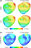
Simulation results from the model of the canine ventricles. (a) Activation maps obtained with the model of the canine ventricles with LDRB and DTI-derived fiber orientations during pacing at the LV epicardium and LV apex. Isochrone lines have a 10 ms spacing. (b) The absolute difference between the activation maps in (a).

References

    1. Alexander AL, Hasan KM, Lazar M, Tsuruda JS, Parker DL. Analysis of partial volume effects in diffusion-tensor MRI. Magn. Reson. Med. 2001;45(5):770–780. - PubMed
    1. Ashikaga H, Criscione JC, Omens JH, Covell JW, Ingels NB. Transmural left ventricular mechanics underlying torsional recoil during relaxation. Am. J. Physiol. Heart Circ. Physiol. 2004;286(2):H640–H647. - PMC - PubMed
    1. Bayer JD, Beaumont J, Krol A. Laplace–Dirichlet energy field specification for deformable models. An FEM approach to active contour fitting. Ann. Biomed. Eng. 2005;33(9):1175–1186. - PubMed
    1. Beyar R, Sideman S. A computer study of the left ventricular performance based on fiber structure, sarcomere dynamics, and transmural electrical propagation velocity. Circ. Res. 1984;55(3):358–375. - PubMed
    1. Bishop MJ, Boyle PM, Plank G, Welsh DG, Vigmond EJ. Modeling the role of the coronary vasculature during external field stimulation. IEEE Trans. Biomed. Eng. 2010;57(10):2335–2345. - PMC - PubMed

Publication types

LinkOut - more resources