. 2012 Dec;3(4):265-75.
doi: 10.1007/s13539-012-0071-9. Epub 2012 May 31.

Cancer cachexia is associated with a decrease in skeletal muscle mitochondrial oxidative capacities without alteration of ATP production efficiency

Affiliations

Cancer cachexia is associated with a decrease in skeletal muscle mitochondrial oxidative capacities without alteration of ATP production efficiency

Cloé M Julienne et al. J Cachexia Sarcopenia Muscle. 2012 Dec.

Abstract

Background: Cancer cachexia is a complex syndrome related to a negative energy balance resulting in muscle wasting. Implication of muscle mitochondrial bioenergetics alterations during cancer cachexia was suggested. Therefore, the aim of this study was to explore the efficiency of oxidative phosphorylation in skeletal muscle mitochondria in a preclinical model of cancer cachexia.

Methods: Berlin-Druckrey IX rats with peritoneal carcinosis (PC) were used as a model of cancer cachexia with healthy pair-fed rats (PF) as control. Hindlimb muscle morphology and fibre type composition were analysed in parallel with ubiquitin ligases and UCP gene expression. Oxidative phosphorylation was investigated in isolated muscle mitochondria by measuring oxygen consumption and ATP synthesis rate.

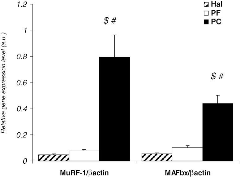
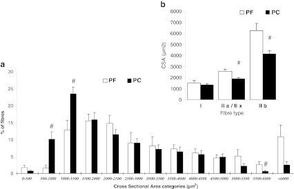
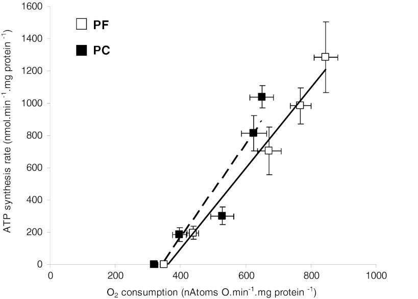
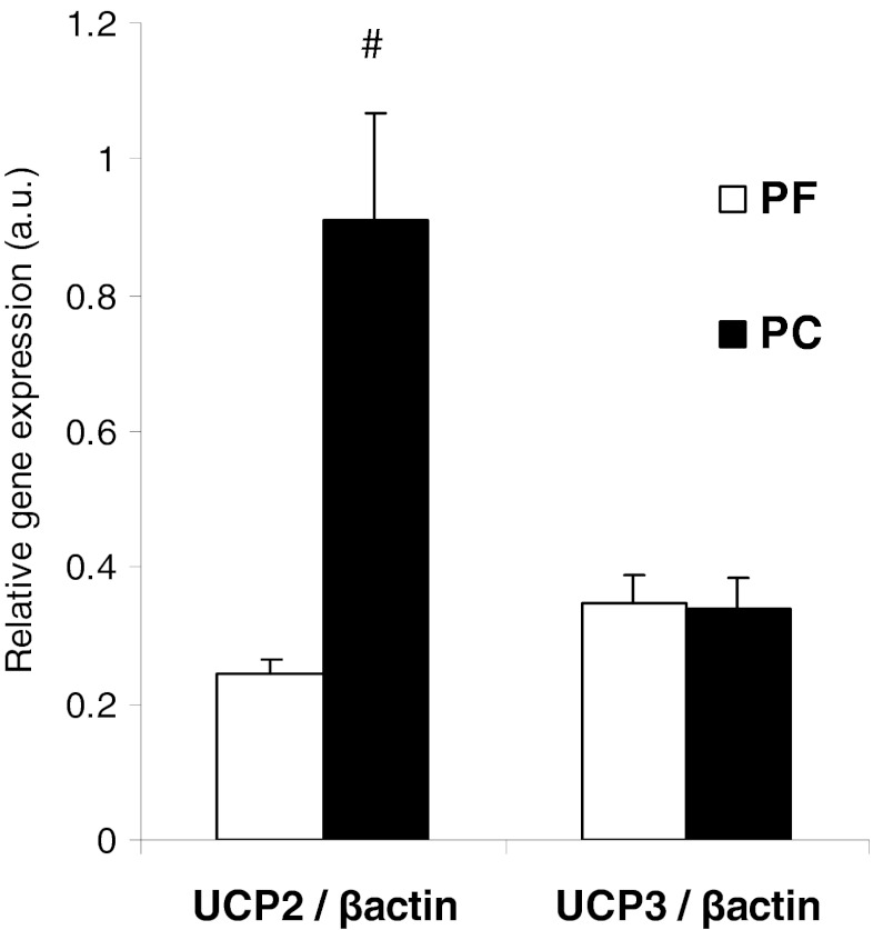
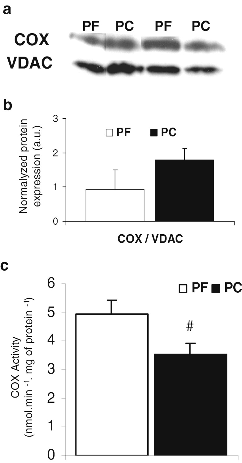
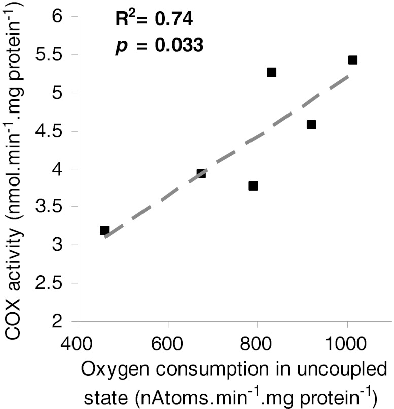
Results: PC rats underwent significant muscle wasting affecting fast glycolytic muscles due to a reduction in fibre cross-sectional area. MuRF1 and MAFbx gene expression were significantly increased (9- and 3.5-fold, respectively) in the muscle of PC compared to PF rats. Oxygen consumption in non-phosphorylating state and the ATP/O were similar in both groups. Muscle UCP2 gene was overexpressed in PC rats. State III and the uncoupled state were significantly lower in muscle mitochondria from PC rats with a parallel reduction in complex IV activity (-30 %).

Conclusion: This study demonstrated that there was neither alteration in ATP synthesis efficiency nor mitochondrial uncoupling in skeletal muscle of cachectic rats despite UCP2 gene overexpression. Muscle mitochondrial oxidative capacities were reduced due to a decrease in complex IV activity. This mitochondrial bioenergetics alteration could participate to insulin resistance, lipid droplet accumulation and lactate production.


Figures

Fig. 1
Fig. 1
Proteolysis in quadriceps muscle from peritoneal carcinosis (PC), pair-fed (PF) and healthy ad libitum (Hal) rats. MuRF1 and MAFbx gene expression, with PF (n = 10) and PC (n = 10). Gene expression levels were normalised for β-actin mRNA. Values are means ± SEM. Statistical significance was set at p < 0.05. Number sign, significantly different from PF. Dollar sign, significantly different from Hal
Fig. 2
Fig. 2
Distribution of quadriceps fibres according to cross-sectional area (CSA) and mean cross-sectional area of type I, IIa/IIx and IIb fibres in pair-fed (PF) and peritoneal carcinosis (PC) rats. a Distribution of quadriceps fibres and b mean cross-sectional area. CSA were determined on approximately 150 fibres in two random fields in PF rats (n = 4) and PC rats (n = 7). Values are means ± SEM. Statistical significance was set at p < 0.05. Number sign, significantly different from PF
Fig. 3
Fig. 3
Efficiency of ATP synthesis in isolated quadriceps mitochondria from pair-fed (PF) and peritoneal carcinosis (PC) rats. ATP/O: relationship between oxygen consumption and ATP synthesis rate. With PF (n = 10) and PC (n = 10), ATP synthesis was measured by the addition of increasing hexokinase concentration. Values are means ± SEM
Fig. 4
Fig. 4
UCP2 and UCP3 gene expression in isolated quadriceps mitochondria from pair-fed (PF) and peritoneal carcinosis (PC) rats. With PF (n = 8) and PC (n = 8). Values are means ± SEM. Statistical significance was set at p < 0.05. Number sign, significantly different from PF
Fig. 5
Fig. 5
Cytochrome c oxidase (COX) protein expression and activity in isolated quadriceps mitochondria from pair-fed (PF) and peritoneal carcinosis (PC) rats. a Western blots of COX protein expression, b Quantification using Quantity One software (Bio-Rad) of the ratio COX/VDAC protein expression. With PF (n = 6) and PC (n = 6), c Enzymatic activity of COX. With PF (n = 4) and PC (n = 4). Values are means ± SEM. Statistical significance was set at p < 0.05. Number sign, significantly different from PF
Fig. 6
Fig. 6
Relationship between cytochrome c oxidase activity and maximal oxygen consumption in uncoupled state in quadriceps isolated mitochondria. The correlation was tested by non-parametric Spearman test

References

    1. Tisdale MJ. Mechanisms of cancer cachexia. Physiol Rev. 2009;89:381–410. doi: 10.1152/physrev.00016.2008. - DOI - PubMed
    1. Acharyya S, Butchbach ME, Sahenk Z, Wang H, Saji M, Carathers M, et al. Dystrophin glycoprotein complex dysfunction: a regulatory link between muscular dystrophy and cancer cachexia. Cancer Cell. 2005;8:421–32. doi: 10.1016/j.ccr.2005.10.004. - DOI - PubMed
    1. Marin-Corral J, Fontes CC, Pascual-Guardia S, Sanchez F, Olivan M, Argiles JM, et al. Redox balance and carbonylated proteins in limb and heart muscles of cachectic rats. Antioxid Redox Signal. 2010;12:365–80. doi: 10.1089/ars.2009.2818. - DOI - PubMed
    1. Argiles JM, Lopez-Soriano FJ, Busquets S. Mechanisms to explain wasting of muscle and fat in cancer cachexia. Curr Opin Support Palliat Care. 2007;1:293–8. doi: 10.1097/SPC.0b013e3282f34738. - DOI - PubMed
    1. Khal J, Hine AV, Fearon KC, Dejong CH, Tisdale MJ. Increased expression of proteasome subunits in skeletal muscle of cancer patients with weight loss. Int J Biochem Cell Biol. 2005;37:2196–206. doi: 10.1016/j.biocel.2004.10.017. - DOI - PubMed

LinkOut - more resources