. 2012 Aug;14(8):1026-36.
doi: 10.1093/neuonc/nos122. Epub 2012 May 30.

VHL regulates the effects of miR-23b on glioma survival and invasion via suppression of HIF-1α/VEGF and β-catenin/Tcf-4 signaling

Affiliations

VHL regulates the effects of miR-23b on glioma survival and invasion via suppression of HIF-1α/VEGF and β-catenin/Tcf-4 signaling

Lingchao Chen et al. Neuro Oncol. 2012 Aug.

Abstract

Aberrant microRNA expression has been implicated in the development of human cancers. Here, we investigated the oncogenic significance and function of miR-23b in glioma. We identified that the expression of miR-23b was elevated in both glioma samples and glioma cells, indicated by real-time polymerase chain reaction analyses. Down-regulation of miR-23b triggered growth inhibition, induced apoptosis, and suppressed invasion of glioma in vitro. Luciferase assay and Western blot analysis revealed that VHL is a direct target of miR-23b. Restoring expression of VHL inhibited glioma proliferation and invasion. Mechanistic investigation revealed that miR-23b deletion decreased HIF-1α/VEGF expression and suppressed β-catenin/Tcf-4 transcription activity by targeting VHL. Furthermore, expression of VHL was inversely correlated with miR-23b in glioma samples and was predictive of patient survival in a retrospective analysis. Therefore, we demonstrated that downregulation of miR-23b suppressed tumor survival through targeting VHL, leading to the inhibition of β-catenin/Tcf-4 and HIF-1α/VEGF signaling pathways.


Figures

Fig. 1.
Fig. 1.
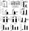
Altered expression of miR-23b in glioma and glioma cell lines. (A) MiR-23b expression in glioma and normal brain tissues by qRT-PCR. (B) qRT-PCR analysis shows that LN229, U87 and U251 glioma cells express higher levels of miR-23b, compared with H4 cell line. Data represent mean ± standard deviation of 3 replicates. *P < .05.
Fig. 2.
Fig. 2.
As-miR-23b suppresses glioma cell proliferation and invasion and induces apoptosis in vitro. (A) miR-23b expression was quantified by qRT-PCR analysis. As-miR-23b significantly reduced miR-23b expression (normalized to U6 RNA) by 89% in U87 cells (P < .01) and 79% in LN229 cells (P < .01), compared with the scramble. (B) Representative histogram showing the total numbers of colonies formed by As-miR-23b treated cells, standardized against scramble cells (normalized to 100%). (C) and (D) Representative images of in vitro cell cycle and transwell assays of U87 and LN2291 after transfected with As-miR-23b or scramble oligonucleotides. (E) and (F) Annexin V-PI and caspase 3/7 activity assays indicate increased apoptosis in U87 and LN229 cells following As-miR-23b treatment. Data represent mean ± standard deviation of 3 replicates. *P < .05.
Fig. 3.
Fig. 3.
VHL is a direct target of miR-23b in glioma cells. (A) Diagram of seed sequence of miR-23b matched the 3′ UTR of the VHL gene and the design of wild or mutant VHL 3′UTR containing reporter constructs. (B) Western blot for VHL expression 48 h after transfected with As-miR-23b or miR-23b. (C) and (D) Luciferase reporter assays in glioma cells after cotransfection of cells with wild-type or mutant 3′UTR VHL and miRNA. Data represent the fold-change of the expression (mean ± standard error) of 3 replicates.
Fig. 4.
Fig. 4.
VHL impacts proliferation, invasion and apoptosis of glioma cells. (A) and (B) VHL mRNA and protein expression levels in U87 and LN229 cells, when transfected with VHL plasmid, were assessed by real-time PCR and Western blot. (C) Proliferation of U87 and LN229 cells after plasmid transfection was significantly reduced compared with the scramble. (D) A significant decrease was observed in the number of invasive U87 and LN229 cells after VHL transfection compared with the scramble. (F) Cell cycle analysis showed an increase in the G0/G1 phase of U87 and LN229 cells when transfected with VHL. (E) and (G) Apoptosis assay showed induction of apoptosis after transfected with VHL. (H–J) Restoration of VHL expression counteracts the impact of miR-23b on proliferation, invasion and apoptosis in SNB 19 cells. *P < .05.
Fig. 5.
Fig. 5.
MiR-23b abrogation suppresses β-catenin/Tcf-4 transcriptional activity and HIF/VEGF signaling. (A) U87 and LN229 cells were cotransfected with Top/Fop, miR23b inhibitor or VHL. And luciferase reporter assays were performed. (B) and (C) Western blot and immunofluorescence detection of β-catenin 48 h after transfection of U87 and LN229 cells with As-miR-23b or VHL. (D) and (E) ELISA and Western blot detection of VEGF 48 h following transfection of U87 and LN229 cells with As-miR-23b or VHL. (F) Real-time PCR analysis of downstream target genes 48 h following transfection of U87 and LN229 cells with As-miR-23b or VHL.
Fig. 6.
Fig. 6.
Knockdown of miR-23b expression impairs LN229 tumor growth in vivo. (A) Luminescence imaging for As-miR-23b-treated LN229 tumors vs. scramble-treated controls. (B) Representative photomicrographs of tumor sections following FISH analysis for miR-23b and IH analysis for VHL. (C) miR-23b and VHL expression in intracranial graft assessed by real-time PCR and Western blot (D) β-catenin and HIF-1α expression in tumor sections following IH analysis. *P < .05.
Fig. 7.
Fig. 7.
VHL expression inversely correlates with miR-23b expression in glioma. (A) Expression of VHL and miR-23b in resected glioma specimen was assessed by IHC and FISH assay. (B) A statistically significant inverse correlation between miR-23b and VHL scores in clinical specimens (Spearman's correlation analysis, r = −0.72854; P < .001). (C) Kaplan–Meier survival curves indicating patients with low VHL expression experience significantly worse outcome compared with VHL high expressed. (D) Schematic representation of the roles of miR-23b and VHL in regulation of proliferation, apoptosis, angiogenesis and invasion.

Similar articles

Cited by

References

    1. Bartel DP. MicroRNAs: target recognition and regulatory functions. Cell. 2009;136(2):215–233. doi:10.1016/j.cell.2009.01.002. - DOI - PMC - PubMed
    1. Lewis BP, Burge CB, Bartel DP. Conserved seed pairing, often flanked by adenosines, indicates that thousands of human genes are microRNA targets. Cell. 2005;120(1):15–20. doi:10.1016/j.cell.2004.12.035. - DOI - PubMed
    1. Calin GA, Croce CM. MicroRNA signatures in human cancers. Nat Rev Cancer. 2006;6(11):857–866. doi:10.1038/nrc1997. - DOI - PubMed
    1. Bandyopadhyay S, Mitra R, Maulik U, Zhang MQ. Development of the human cancer microRNA network. Silence. 2010;1(1):6. doi:10.1186/1758-907X-1-6. - DOI - PMC - PubMed
    1. Van Meir EG, Hadjipanayis CG, Norden AD, Shu HK, Wen PY, Olson JJ. Exciting new advances in neuro-oncology: the avenue to a cure for malignant glioma. CA Cancer J Clin. 2010;60(3):166–193. doi:10.3322/caac.20069. - DOI - PMC - PubMed

Publication types

MeSH terms

Substances