. 2012 May 30;32(22):7439-53.
doi: 10.1523/JNEUROSCI.5318-11.2012.

Doublecortin (DCX) mediates endocytosis of neurofascin independently of microtubule binding

Affiliations

Doublecortin (DCX) mediates endocytosis of neurofascin independently of microtubule binding

Chan Choo Yap et al. J Neurosci. .

Abstract

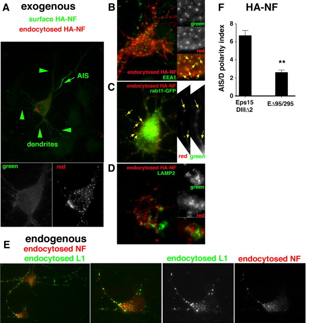
Doublecortin on X chromosome (DCX) is one of two major genetic loci underlying human lissencephaly, a neurodevelopmental disorder with defects in neuronal migration and axon outgrowth. DCX is a microtubule-binding protein, and much work has focused on its microtubule-associated functions. DCX has other reported binding partners, including the cell adhesion molecule neurofascin, but the functional significance of the DCX-neurofascin interaction is not understood. Neurofascin localizes strongly to the axon initial segment in mature neurons, where it plays a role in assembling and maintaining other axon initial segment components. During development, neurofascin likely plays additional roles in axon guidance and in GABAergic synaptogenesis. We show here that DCX can modulate the surface distribution of neurofascin in developing cultured rat neurons and thereby the relative extent of accumulation between the axon initial segment and soma and dendrites. Mechanistically, DCX acts via increasing endocytosis of neurofascin from soma and dendrites. Surprisingly, DCX increases neurofascin endocytosis apparently independently of its microtubule-binding activity. We additionally show that the patient allele DCXG253D still binds microtubules but is deficient in promoting neurofascin endocytosis. We propose that DCX acts as an endocytic adaptor for neurofascin to fine-tune its surface distribution during neuronal development.


Figures

Figure 1.
Figure 1.
Downregulating DCX levels affects neurofascin surface distribution between dendrites and the AIS. A, Cultured neurons (DIV9/10) were transfected with shRandom-GFP (left panels) or shDCX#2-GFP (right panels; blue) for 20 h and then stained against endogenous DCX (red). Single-channel images of the DCX staining are shown in the bottom panels. The transfected cell is indicated with an arrow. B, Mature, cultured neurons were transfected with shRandom-GFP or four different shDCX-GFP plasmids and then stained against endogenous DCX. Levels of endogenous DCX were determined from immunofluorescence images and normalized to untransfected cells. The extent of downregulation was determined after 20 h of shRNA expression in DIV10 cells to parallel experiments using acute HA-NF expression, or after 7–8 d of shRNA expression in DIV14 cells to parallel experiments with endogenous proteins. ***p < 0.0001; **p < 0.001; Bars, SEM. C, Surface HA-NF was stained in cells expressing HA-NF and either shRandom-GFP as control (left panel) or shDCX#2-GFP (right panel) for 20 h. Arrow indicates AIS. D, AIS intensity was determined for endogenous neurofascin (DIV14, plasmid expression for 7 d) after transfection with shRandom, shDCX#2, or shDCX#2 with resistant DCX. N = 110 cells shRandom, 94 cells shDCX#2, 83 cells shDCX#2 plus resistant DCX. ***p < 0.0001; *p < 0.01; Bars, SEM. E, F, Quantification of HA-NF surface staining on AIS versus dendrites (AIS/D) in cells expressing shRandom-GFP, shDCX#2-GFP, or shDCX#2-GFP with an shRNA-resistant DCX (DIV9/10, plasmid expression for 20 h). Scatter plot (E) of dendrite intensity and HA-NF AIS/D polarity index for individual cells transfected with shRandom-GFP (blue) or shDCX (red) for 20 h; AU, Arbitrary units. Average values from all cells are displayed as squares and are graphed in the histogram in (F) for the AIS/D polarity index. N = 113 cells shRandom, 113 cells shDCX#2, 121 cells shDCX#2 + resistant DCX. ***p < 0.0001; Bars, SEM. GI, Effects of DCX downregulation in neurons on two other membrane proteins and the cytoskeletal AIS component ankG were quantified: exogenously expressed NgCAM (expression for 20 h, fixed DIV10) (G), endogenous voltage-gated sodium channel (VGSC) (expression for 7 d, fixed DIV14) (H), and ankyrinG (expression for 7 d, fixed DIV14) (I). NgCAM: n = 33 cells shRandom, n = 25 cells shDCX; VGSC: n = 22 cells shRandom, n = 24 cells shDCX; AnkyrinG: n = 30 cells shRandom, n = 23 cells shDCX.
Figure 2.
Figure 2.
NF and DCX colocalize intracellularly in neurons. A, Costaining of DIV9 neurons with antibodies against extracellular neurofascin (live staining; red) and endogenous DCX (aqua). No co-colocalization is observed at the AIS (arrowheads). B, Costaining of DIV9 neurons with antibodies against intracellular epitope of neurofascin (red) and endogenous DCX (aqua). No co-colocalization is observed at the AIS, but colocalizing puncta can be observed along axons and dendrites (arrows in insets). C, DIV9 neurons were transfected with HA-NF and stained with antibodies against an intracellular epitope of neurofascin in red and endogenous DCX in green. Colocalizing puncta can be observed along axons and dendrites.
Figure 3.
Figure 3.
Neurofascin and DCX reside in close proximity in cultured neurons using Duolink. A, B, Duolink detection was performed on DIV12 hippocampal cultures using cytoplasmic tail-directed NF antibody and DCX antibody (A) or only the cytoplasmic tail-directed NF antibody (B), and Duolink signal was detected in red (single channel shown in right panels). Subsequently, NF antibody was detected with a green secondary, and DCX antibody was detected in blue. Abundant Duolink dots were detected along dendrites in A (A′ shows close-up of one dendrite), but no dots in the control (B). C, D, Hippocampal neurons were transfected at DIV7 with either shRandom-GFP (C) or shDCX#2-GFP (D), and Duolink was performed 5 d later with cytoplasmic tail-directed NF antibody and DCX antibody (as in A). C, Abundant Duolink signal (middle panel) was detected along transfected (green) dendrites, which expressed DCX (blue). Single channels of one transfected dendrite (outlined and marked by green arrows) are shown in the small panels below. D, Few Duolink dots are present on the sh-DCX#2-GFP transfected neuron (green arrow), but abundant Duolink dots are present along dendrites of nontransfected cells, still expressing DCX (blue). Single channels of one transfected dendrite (outlined by green arrows) are shown in the small panels. A nontransfected dendrite crossing the field is outlined with blue and marked with blue arrows and still expresses DCX. Line scans of the Duolink signal along the dendrites corresponding to the small panels are shown in E (shRandom-GFP) and F (shDCX#2-GFP).
Figure 4.
Figure 4.
Neurofascin/DCX complex in vitro. A, Coimmunoprecipitation of DCX and neurofascin from E18 rat brain membrane fraction with nonimmune IgG (lane 1) or anti-neurofascin antibody (lane 2). The levels of neurofascin and DCX in the lysates were determined by Western blotting (WB). IP, Immunoprecipitation B, Presence of phospho-FIGQY neurofascin in hippocampal cultures. Hippocampal cultures at DIV10 were treated with 100 ng/ml NGF for 48 h (lanes 1 and 3) or not (lane 2), and membrane fractions were prepared and used for immunoprecipitation with control IgG (lane 1) or anti-neurofascin (NF) antibody (lanes 2 and 3), and the immunoprecipitates were probed with anti-phosphoFIGQY antibody. PhosphoFIGQY could be detected even without additional NGF, but NGF treatment increased the levels of phosphoFIGQY. MW, Molecular weight. C, Diagram of DCX-GFP indicating the DC repeats and the positions of the G253 and R89 patient mutations. D, HEK293 cells were transfected with HA-NF and GFP (lane 1), DCX-GFP (lane 2), or DCX-G253D-GFP (lane 3). Lysates were prepared and used for WB against HA or GFP or used for IP with anti-HA antibody. DCX-GFP was efficiently immunoprecipitated, whereas DCX-G253D-GFP was not (arrow).
Figure 5.
Figure 5.
Coexpression of DCX mutants deficient for NF binding interferes with HA-NF surface distribution. A, B, Scatter plots of dendrite intensity and AIS/D polarity index for individual cells transfected with GFP (blue) or DCX-GFP (red) (B) for 20 h, or with GFP (blue) or DCX-G253D-GFP (red) (B). Average (avg) values are displayed as squares and are graphed in the histogram in F for the AIS/D polarity index. CE, Surface HA-NF (red) was stained after transfection with HA-NF and either GFP (C), DCX-GFP (D), or DCX-G253D-GFP (E). Single channels of surface HA-NF are shown in the right panels. F, Overexpression of DCX-GFP for 20 h does not change AIS/D polarity index. DCX-G253D-GFP decreases relative AIS targeting of HA-NF. N = 139 cells for GFP, 119 cells for DCX-GFP, and 136 cells for DCX-G253D from three independent experiments. ***p < 0.0001; Bars, SEM.
Figure 6.
Figure 6.
DCX-G253D mutant still binds microtubules in cells. AB′, Neuronal cultures were transfected with DCX-GFP (A, A′) or DCX-G253D-GFP (B, B′). After 20 h, live cultures were either fixed (A, B) or detergent extracted in microtubule-stabilizing buffer BRB80 before fixation (A′, B′). The transfected proteins were detected with anti-GFP (green) antibodies and counterstained for MAP2 (red). Both constructs expressed well in transfected neurons without extractions. If cells were extracted live before fixation, both DCX constructs were still easily detected on detergent-insoluble cytoskeleton. C, COS cells extracted in BRB80 and 0.15% Triton X-100 before fixation show intact microtubule (blue) and microfilament (red) networks. D, COS cells extracted in PBS++ and 0.15% Triton X-100 before fixation show intact microfilament network (red), but loss of microtubules (blue). EG, COS cells were transfected with DCX-GFP (E, F) or DCX-G253D-GFP (G) and extracted in BRB80 (E, G) or microtubule-destabilizing buffer PBS++ (F). DCX-GFP decorates individual microtubules (blue) (Ea) and induces microtubule bundles when highly expressed (Eb). After extraction in PBS++, DCX is nondetectable in transfected cells (expressing lamin-cherry) (F). DCX-G253D-GFP also associates with microtubules and still induces bundles (G).
Figure 7.
Figure 7.
Overexpression of a microtubule-binding deficient DCX (DCX-R89G) does not cause mislocalization of HA-NF. A, Expression levels of DCX-R89G-GFP were determined in lysates of HEK293 cells coexpressing HA-NF and either DCX-GFP or DCX-R89G-GFP. HA-NF was used for normalization of transfection efficiency and loading. MW, Molecular weight. B, HEK293 cells were transfected with HA-NF and GFP (lane 1), DCX-GFP (lane 2), or DCX-R89G-GFP (lane 3). Lysates were prepared and used for Western blotting (WB) against HA or GFP or used for immunoprecipitation (IP) with anti-HA antibody. DCX-GFP was efficiently immunoprecipitated (top panel, arrow). DCX-R89G-GFP was expressed at much reduced levels in the lysate, but could still be detected as a coimmunoprecipitating (co-IP) band (lane 3; arrow). CE, DCX-R89G-GFP has reduced microtubule-binding capacity. C, COS cells were transfected with GFP, DCX-R89G-GFP, or DCX-GFP, fixed, and stained with anti-GFP antibody. GFP and DCX-R89G-GFP are cytosolic and are not associated with microtubules, whereas DCX-GFP decorates microtubules. D, E, COS cells transfected with DCX-GFP (D) or DCX-R89G-GFP (E) were extracted in BRB80. DCX-GFP (green) localizes along microtubules (red) (D). DCX-R89G-GFP, on the other hand, is not detectable (green) despite the presence of microtubules (blue) (E). Lamin-cherry was cotransfected (red) with DCX-R89G-GFP to mark transfected cells. F, Expression of DCX-R89G-GFP in hippocampal neurons does not lead to mislocalization of surface HA-NF (red). A representative experiment is shown (of three independent experiments). N = 63 cells for GFP, 57 cells for DCX-GFP, and 77 cells for DCX-R89G-GFP. Bars, SEM. The differences are not statistically significant.
Figure 8.
Figure 8.
Neurofascin undergoes endocytosis in hippocampal neurons. A, HA-NF can be found in endosomes (red), especially in the somatodendritic domain after 30 min of rabbit anti-HA antibody uptake. Surface HA-NF (green) was stained with mouse anti-HA antibody before fixation and permeabilization. A zoom image of the soma region is shown in the bottom panels, with each channel shown separately. BD, Costaining of endocytosed HA-NF (red) with markers (green) to early endosomes (EEA1) (B), recycling endosomes (rab11-GFP) (C), and late endosomes/lysosomes (LAMP2) after 90 minute chase D, Zoomed-in regions are shown as single channels in the small panels. Yellow arrows point at colocalizing puncta. E, DIV3 hippocampal neurons were incubated live with antibodies against extracellular domains of NF (red) and L1 (green) for 30 min. Both L1 and NF can be detected in endosomes in the soma as well as along processes. One cell is shown with single channels. F, Dominant-negative Eps15 construct (EΔ95/295-GFP) and an inactive Eps15 construct (Eps15DIIIΔ2-GFP) were expressed together with HA-NF for 20 h, and AIS/D polarity index was determined. Inhibition of clathrin-mediated endocytosis with EΔ95/295-GFP decreased AIS localization compared to controls (Eps15DIIID2-GFP). N = 15–30 cells per experiment and condition in three independent experiments. **p < 0.001; Bars, SEM.
Figure 9.
Figure 9.
Endocytosis of HA-NF is stimulated by coexpression of DCX-GFP in PC12 cells. A, PC12 cells were transfected with HA-NF and GFP (A, top panels) or DCX-GFP (A, bottom panels) in the presence of NGF and endocytosis assays carried out with anti-HA-antibody. Individual channels for a representative cell are shown. B, C, Quantification of internalized HA-NF. Two to five independent experiments were analyzed. Results from one representative experiment are shown. B, N = 59 cells for controls, 82 cells for DCX-GFP, 84 cells for DCX-G253D-GFP. C, N = 76 cells for controls, 91 cells for DCX-GFP, 84 cells for DCX-R89G-GFP; ***p < 0.0001; **p < 0.001. D, DCX-GFP does not promote the endocytosis of L1-myc. N = 34 cells for GFP, 32 cells for DCX-GFP. The differences are not statistically significant. E, DCX-GFP does not promote the endocytosis of transferrin. N = 72 cells for GFP, 76 cells for DCX-GFP. The differences are not statistically significant. Bars, SEM.
Figure 10.
Figure 10.
Endocytosis of endogenous NF is diminished after downregulation of DCX in neurons. A, B, Dissociated hippocampal neurons were electroporated with either shRandom-GFP (A) or shDCX#2-GFP (B) before plating and endocytosis assays with anti-NF antibody carried out at DIV3. Cultures were fixed and stained against GFP (green), DCX (blue), and endocytosed anti-NF (red). The arrow points at the transfected cell. DCX (blue) is diminished in the cell expressing shDCX#2-GFP (B, middle panel), but not in the cell expressing shRandom-GFP (A, middle panel). Red channel alone is shown in the right panel. The cell expressing shDCX#2-GFP (B, right panel) shows less endocytosed NF than the untransfected cells in the same field. C, The levels of endocytosed NF were quantified in cells expressing shDCX#2 compared to untransfected cells (UT) in the same field, or expressing shRandom compared to UT cells. One representative experiment (of two independent experiments) is shown. N = 25 cells for shDCX#2 and 52 for corresponding UT cells; N = 24 cells for shRandom and 26 for corresponding UT cells; ***p < 0.0001. Bars, SEM. D, AIS intensity was determined for endogenous neurofascin in developing neurons (DIV7, plasmid expression for 7 d) after transfection with shRandom or shDCX#2. N = 125 cells for shRandom, n = 126 cells for shDCX#2; ***p < 0.0001; Bars, SEM.

References

    1. Ango F, di Cristo G, Higashiyama H, Bennett V, Wu P, Huang ZJ. Ankyrin-based subcellular gradient of neurofascin, an immunoglobulin family protein, directs GABAergic innervation at purkinje axon initial segment. Cell. 2004;119:257–272. - PubMed
    1. Bai J, Ramos RL, Ackman JB, Thomas AM, Lee RV, LoTurco JJ. RNAi reveals doublecortin is required for radial migration in rat neocortex. Nat Neurosci. 2003;6:1277–1283. - PubMed
    1. Benmerah A, Bayrou M, Cerf-Bensussan N, Dautry-Varsat A. Inhibition of clathrin-coated pit assembly by an Eps15 mutant. J Cell Sci. 1999;112:1303–1311. - PubMed
    1. Boiko T, Vakulenko M, Ewers H, Yap CC, Norden C, Winckler B. Ankyrin-dependent and -independent mechanisms orchestrate axonal compartmentalization of L1 family members neurofascin and L1/neuron-glia cell adhesion molecule. J Neurosci. 2007;27:590–603. - PMC - PubMed
    1. Bréchet A, Fache MP, Brachet A, Ferracci G, Baude A, Irondelle M, Pereira S, Leterrier C, Dargent B. Protein kinase CK2 contributes to the organization of sodium channels in axonal membranes by regulating their interactions with ankyrin G. J Cell Biol. 2008;183:1101–1114. - PMC - PubMed

MeSH terms

LinkOut - more resources