. 2011 Nov 11:2:71.
doi: 10.3389/fendo.2011.00071. eCollection 2011.

Extracellular domain N-glycosylation controls human thrombopoietin receptor cell surface levels

Affiliations

Extracellular domain N-glycosylation controls human thrombopoietin receptor cell surface levels

Roxana I Albu et al. Front Endocrinol (Lausanne). .

Abstract

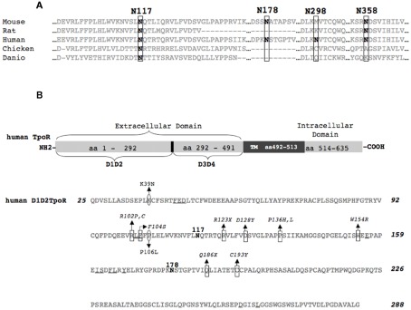
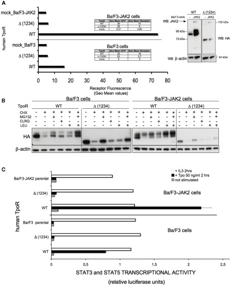
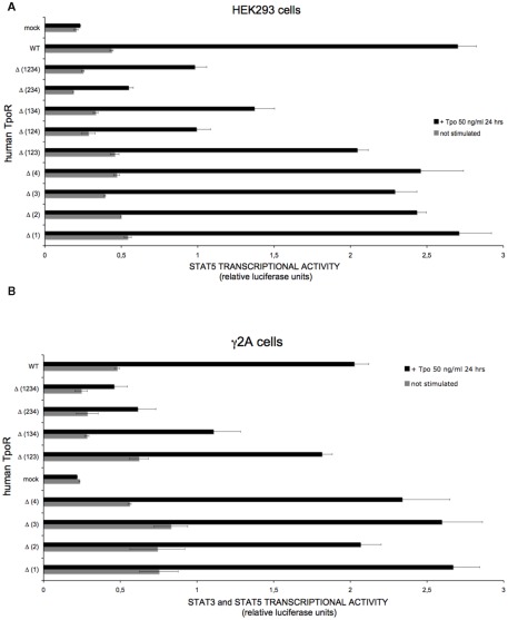
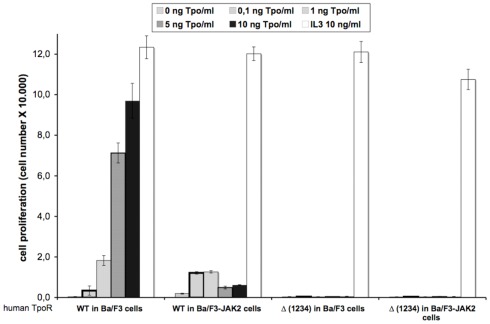
The thrombopoietin receptor (TpoR) is a type I transmembrane protein that mediates the signaling functions of thrombopoietin (Tpo) in regulating megakaryocyte differentiation, platelet formation, and hematopoietic stem cell renewal. We probed the role of each of the four extracellular domain putative N-glycosylation sites for cell surface localization and function of the receptor. Single N-glycosylation mutants at any of the four sites were able to acquire the mature N-glycosylated pattern, but exhibited a decreased Tpo-dependent JAK2-STAT response in stably transduced Ba/F3 or Ba/F3-JAK2 cell lines. The ability of JAK2 to promote cell surface localization and stability of TpoR required the first N-glycosylation site (Asn117). In contrast, the third N-glycosylation site (Asn298) decreased receptor maturation and stability. TpoR mutants lacking three N-glycosylation sites were defective in maturation, but N-glycosylation on the single remaining site could be detected by sensitivity to PNGaseF. The TpoR mutant defective in all four N-glycosylation sites was severely impaired in plasma membrane localization and was degraded by the proteasome. N-glycosylation receptor mutants are not misfolded as, once localized on the cell surface in overexpression conditions, they can bind and respond to Tpo. Our data indicate that extracellular domain N-glycosylation sites regulate in a combinatorial manner cell surface localization of TpoR. We discuss how mutations around TpoR N-glycosylation sites might contribute to inefficient receptor traffic and disease.

Keywords: ER maturation; JAK2; N-glycosylation; cell surface traffic; cytokine receptor; endoglycosidase H; signal transduction; thrombopoietin.


Figures

Figure 1
Figure 1
Schematic representation of human TpoR and multiple sequence alignment of the N-glycosylation sites of TpoR from different species. (A) Multiple sequence alignment showing conservation of the first N-glycosylation site from different species. The alignment was obtained with the ClustalW program for the following sequences: Homo sapiens (UniProtKB/Swiss-Prot accession number P40238), Mus musculus (Q08351), Rattus norvegicus (D4A2R0), Gallus gallus (Q6IYE8), Danio rerio (Q6EIY6). The positions of the four putative N-glycosylation sites of human TpoR are depicted. (B) Schematic representation of human TpoR. The bottom panel of (B) depicts the D1D2 cytokine receptor module. The important aminoacids for Tpo binding are underlined (Deane et al., 1997). In square (□) are depicted the aminoacids that are mutated in congenital amegakaryocytic thrombocytopenia (Ballmaier et al., ; Germeshausen et al., ; Tijssen et al., ; Fox et al., 2009) and in round circles (○) the aminoacids that are mutated in familial thrombocytosis (Moliterno et al., ; El-Harith El et al., 2009). TM, transmembrane region; aa, aminoacid; D1D2, sub-domains of the distal cytokine receptor module; D3D4, sub-domains of the proximal cytokine receptor module; NH2, N-terminus of the protein; COOH, C-terminus of the protein; X, any aminoacid.
Figure 2
Figure 2
Effect of single N-glycosylation site removal on the cell surface levels and stability of human TpoR. (A) Sorted Ba/F3 parental or Ba/F3-JAK2 cells, expressing TpoR variants defective in one N-glycosylation site [Δ (1), Δ (2), Δ (3), Δ (4)] were tested for cell surface levels using anti-HA antibody based flow cytometry. Similar results were obtained in four independent experiments, on two different cell lines obtained for each TpoR variant. The differences between the cell surface levels of Δ (1) TpoR in Ba/F3 and Ba/F3-JAK2 cells were statistically significant when compared with the levels of WT TpoR (p = 0.006 and p = 0.0086, respectively). (B) HEK293 cells were plated in 6-well plates and transiently transfected with 3 μg of each of the indicated TpoR variants. Twenty-four hours post-transfection cells were lysed and treated with EndoH or PNGaseF or left untreated and subjected to Western blot analysis. Membranes were probed with anti-HA. Similar results were obtained in three different experiments. (C) A pool of the cell lines used in (A) were treated at 37°C with 50 μg/ml cycloheximide (CHX) over different time periods then lysed. The lysates were analyzed for TpoR variants stability using anti-HA and normalizing by anti-β-actin Western blotting.
Figure 3
Figure 3
Effect of single N-glycosylation site removal on the Tpo-dependent transcriptional activation of human TpoR. (A) Ba/F3 or Ba/F3-JAK2 cells expressing stably expressing each human TpoR defective in single N-glycosylation site [Δ (1), Δ (2), Δ (3), Δ (4)] were starved 3 h in RPMI medium +1 mg/ml BSA and electroporated with the luciferase reporters pGRR5-luc and pRLTK-luc. The cells were stimulated with 50 ng Tpo/ml or left unstimulated for 2 h. Upon treatment the cells were lysed and their luminescence recorded. Results are the mean ± variation of triplicate samples. One of three independent experiments is depicted. The t-test showed statistically significant differences for the Tpo-dependent transcriptional levels of the TpoR mutants when compared with the WT TpoR (p < 0.05 for all the TpoR variants). Of importance, when Tpo-dependent Δ (1) TpoR transcriptional activity in the stimulated condition was compared with the transcriptional activity of the stimulated WT TpoR, the p value equaled 0.0086. (B) Ba/F3 stably transduced cells for each of the TpoR variants [Δ (1), Δ (2), Δ (3), and Δ (4)] were washed three times and seeded in 96-well plates at a density of 3,000 cells/well. The cells were stimulated with 5 ng Tpo/ml, or with 5 ng IL3/ml or left unstimulated during 72 h. After 72 h, the proliferation of the cells was determined by the DNA synthesis assay reflected by the measurements of the thymidine incorporation as described in Experimental procedures. Results are the mean ± variation of triplicate samples. One of three independent experiments is depicted. The t-test showed significant differences between the Tpo-dependent proliferations level of Δ (1) TpoR (p = 0.0001) and Δ (2) TpoR (p = 0.0019) when compared with WT TpoR.
Figure 4
Figure 4
Effect of multiple N-glycosylation sites removal on the cell surface levels and function of human TpoR. (A) Sorted Ba/F3 parental or Ba/F3-JAK2 cells, expressing TpoR variants defective in three N-glycosylation sites [Δ (123), Δ (124), Δ (134), Δ (234)] were tested for cell surface levels using anti-HA antibody based flow cytometry. Similar results were obtained in two independent experiments, on two different cell lines obtained for each TpoR variant. For the Ba/F3 cells, the t-test showed statistically significant differences between Δ (123) TpoR (p = 0.0019), Δ (124) TpoR (p = 0.0036), Δ (134) TpoR (p = 0.0016), and Δ (234) TpoR (p = 0.0086) when compared with WT TpoR. (B) HEK293 cells were seeded in 6-well plates and transiently transfected with 3 μg of each of the indicated TpoR variants. Twenty-four hours post-transfection cells were lysed and treated with EndoH or PNGaseF or left untreated during 16 h at 37°C. The digestion products were subjected to Western blot analysis. Membranes were probed with anti-HA and anti-β-actin. Similar results were obtained in two different experiments. (C) A pool of each cell line used in (A) was treated at 37°C with 50 μg/ml cycloheximide (CHX) over different time periods then lysed. The lysates were analyzed for TpoR variants stability using anti-HA and normalized by anti-β-actin Western blotting. (D) Ba/F3 or Ba/F3-JAK2 cells stably expressing each human TpoR defective in three N-glycosylation sites were starved 3 h in RPMI medium +1 mg/ml BSA and electroporated with the pGRR5-luc and pRLTK-luc reporters. The cells were stimulated with 50 ng Tpo/ml or left unstimulated 2 h at 37°C. Upon treatment, the cells were lysed and their luminescence recorded. Results are the mean ± variation of triplicate samples. One of three independent experiments is depicted. The t-test showed statistically significant differences for Tpo-dependent transcriptional activity levels of the all the TpoR variants, when compared with the WT TpoR (all the p values < 0.0001).
Figure 5
Figure 5
Removal of all four N-glycosylation sites dramatically affects the cell surface localization of human TpoR in Ba/F3 cells. (A) Ba/F3 or Ba/F3-JAK2 cells stably expressing the WT TpoR or the variant of the receptor defective in all four N-glycosylation sites [Δ (1234)] were tested for cell surface levels using anti-HA antibody based flow cytometry. Similar results were obtained in five independent experiments, on two different cell lines obtained for the Δ (1234) variant (p < 0.0001). Right panel of the (A) shows a Western blot of the cells used in the experiment, probing the receptor with anti-HA antibodies, the murine JAK2 WT with anti-JAK2 antibodies and using anti-β-actin as a control. (B) Ba/F3 or Ba/F3-JAK2 cells used in (A) were tested for the contribution of the proteasome- or lysosome-mediated degradation of the TpoR N-glycosylation defective mutant Δ (1234) versus WT. The cells were incubated 3 h at 37°C with 50 μg/ml cycloheximide and proteasome inhibitor MG132 (10 μM) or with 50 μg/ml cycloheximide and lysosome inhibitors (200 μM chloroquine or 10 μg/ml leupeptin). Control cells were kept untreated to have 100% protein expression. Cells were lysed and analyzed by Western blotting with antibodies against HA for the levels of TpoR and anti-β-actin as a loading control. (C) Pools from the same cell lines used in (A,B) were starved 3 h in RPMI medium +1 mg/ml BSA and electroporated with pGRR5-luc and pRLTK-luc reporters. The cells were also stimulated with Tpo or left unstimulated during 2 h at 37°C. Upon treatment the cells were lysed and their luminescence recorded. Results are the mean ± variation of triplicate samples. One of three independent experiments is depicted. The t-test showed statistically significant differences between the Tpo-dependent transcriptional activities level of the Δ (1234) TpoR and that of WT TpoR (p < 0.0001 for both Ba/F3 and Ba/F3-JAK2 cell lines).
Figure 6
Figure 6
Human TpoR mutants defective in N-glycosylation sites are able, in overexpression conditions, to support Tpo-dependent STAT3 and STAT5 transcriptional activities. (A) HEK293 cells were plated in 24-well plates the day before transfection. The cells were co-transfected with plasmids encoding for each of the TpoR variants, the pLHRE-luc and pRLTK-luc reporters, and JAK2 WT. Cells were lysed 24 h after treatment in 100 μl 1× passive lysis buffer and luminescence values were recorded. The experiment was independently performed three times and the presented result represents ± variation of triplicate samples. The t-test showed that the unstimulated condition is significantly different from the stimulated condition for the Δ (1234) TpoR (p = 0.0058). (B), JAK2-deficient γ2A cells were plated in 24-well plates a day before transfection. The cells were co-transfected with plasmids encoding for each of the TpoR variants, the pGRR5-luc and pRLTK-luc reporters, and JAK2 WT. Four hours post-transfection cells were stimulated or left unstimulated. Cells were lysed 24 h after treatment in 100 μl 1× passive lysis buffer and luminescence values were recorded. The experiment was independently performed two times and the result presented represents ± variation of triplicate samples.
Figure A1
Figure A1
Removal of all four N-glycosylation sites dramatically affects the Tpo-dependent cell proliferation of human TpoR in Ba/F3 and Ba/F3-JAK2 cells. Ba/F3 and Ba/F3-JAK2 cells stably expressing WT or Δ (1234) human TpoR were washed 3 times and seeded in 96-well plates at a density of 3,000 cells/well. The cells were stimulated with different Tpo concentrations, or with 5 ng IL3/ml or left unstimulated during 72 h. After 72 h, cell proliferation was determined by the DNA synthesis assay reflected by the measurements of the thymidine incorporation as described in Experimental procedures. Results are the mean ± variation of triplicate samples. One of two independent experiments is depicted.

References

    1. Ballmaier M., Germeshausen M., Schulze H., Cherkaoui K., Lang S., Gaudig A., Krukemeier S., Eilers M., Strauss G., Welte K. (2001). c-mpl Mutations are the cause of congenital amegakaryocytic thrombocytopenia. Blood 97, 139–14610.1182/blood.V97.1.139 - DOI - PubMed
    1. Bazan J. F. (1990). Structural design and molecular evolution of a cytokine receptor superfamily. Proc. Natl. Acad. Sci. U.S.A. 87, 6934–693810.1073/pnas.87.18.6934 - DOI - PMC - PubMed
    1. Bolander F. F., Jr. (1999). Regulation of prolactin receptor glycosylation and its role in receptor location. Mol. Cell. Endocrinol. 149, 85–9210.1016/S0303-7207(98)00251-2 - DOI - PubMed
    1. Buteau H., Pezet A., Ferrag F., Perrot-Applanat M., Kelly P. A., Edery M. (1998). N-glycosylation of the prolactin receptor is not required for activation of gene transcription but is crucial for its cell surface targeting. Mol. Endocrinol. 12, 544–55510.1210/me.12.4.544 - DOI - PubMed
    1. Dalal I., Arpaia E., Dadi H., Kulkarni S., Squire J., Roifman C. M. (1998). Cloning and characterization of the human homolog of mouse Jak2. Blood 91, 844–851 - PubMed

LinkOut - more resources