. 2012;7(5):e36941.
doi: 10.1371/journal.pone.0036941. Epub 2012 May 23.

Suppression of AP1 transcription factor function in keratinocyte suppresses differentiation

Affiliations

Suppression of AP1 transcription factor function in keratinocyte suppresses differentiation

Bingshe Han et al. PLoS One. 2012.

Retraction in

Abstract

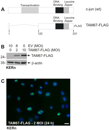
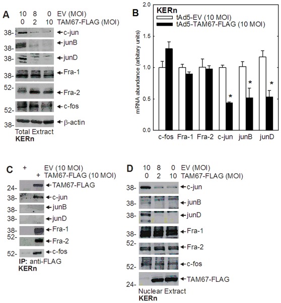
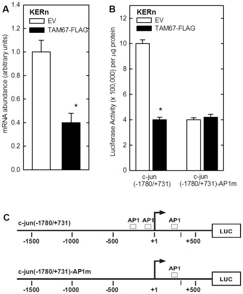
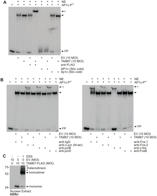
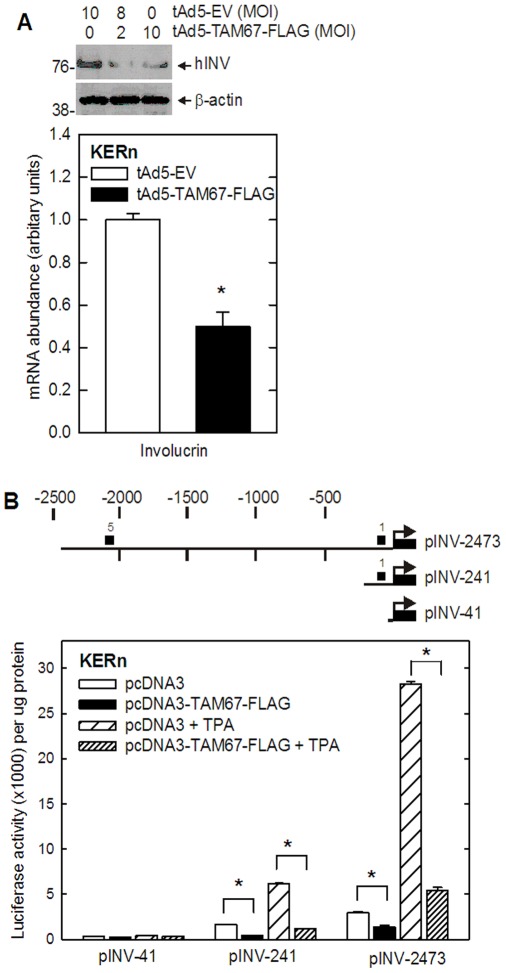
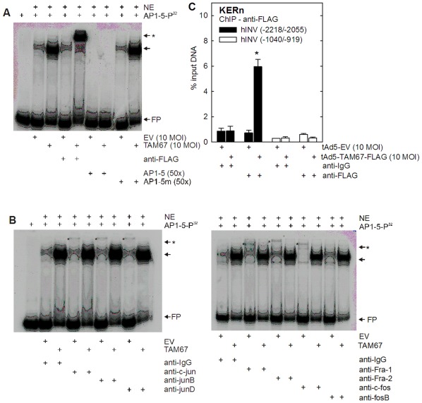
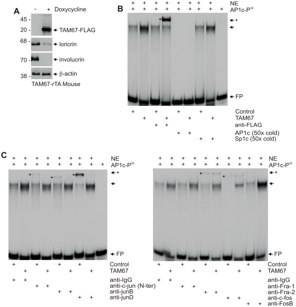
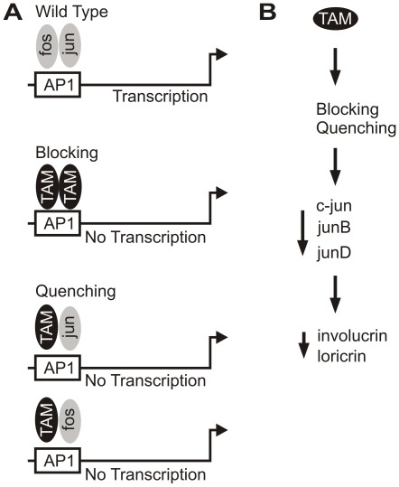
Our previous study shows that inhibiting activator protein one (AP1) transcription factor function in murine epidermis, using dominant-negative c-jun (TAM67), increases cell proliferation and delays differentiation. To understand the mechanism of action, we compare TAM67 impact in mouse epidermis and in cultured normal human keratinocytes. We show that TAM67 localizes in the nucleus where it forms TAM67 homodimers that competitively interact with AP1 transcription factor DNA binding sites to reduce endogenous jun and fos factor binding. Involucrin is a marker of keratinocyte differentiation that is expressed in the suprabasal epidermis and this expression requires AP1 factor interaction at the AP1-5 site in the promoter. TAM67 interacts competitively at this site to reduce involucrin expression. TAM67 also reduces endogenous c-jun, junB and junD mRNA and protein level. Studies with c-jun promoter suggest that this is due to reduced transcription of the c-jun gene. We propose that TAM67 suppresses keratinocyte differentiation by interfering with endogenous AP1 factor binding to regulator elements in differentiation-associated target genes, and by reducing endogenous c-jun factor expression.


Conflict of interest statement

Competing Interests: Richard L. Eckert is a PloS ONE Editor Board member, but this does not alter his adherence to all of the PloS ONE policies on sharing data and materials, as detailed online in the guide for authors. The other authors have declared that no competing interests exist.

Figures

Figure 1
Figure 1. TAM67-FLAG expression in keratinocytes. A
Comparison of c-jun and TAM67 structure. The numbers are indicated in amino acids. The transactivation, DNA binding and leucine zipper domains are indicated. The TAM67 truncated protein is FLAG epitope tagged as indicated. B/C TAM67-FLAG is expressed in keratinocytes. Normal human keratinocytes were infected with 10 MOI of tAd5-EV or tAd5-TAM67-FLAG with 5 MOI of Ad5-TA. After 24 h the cells were fixed for immunostaining and extracts were prepared for immunoblot with anti-FLAG. Similar results were observed in each of three repeated experiments.
Figure 2
Figure 2. Impact of TAM67-FLAG on AP1 factors.
Keratinocytes were infected with empty (EV) or TAM67-FLAG encoding adenovirus and after 24 h cells were harvested and extracts prepared. A Total extracts were electrophoresed for immunoblot detection of the indicated proteins. B TAM67-FLAG suppresses jun factor mRNA level. At 24 h post-infection with EV or TAM67-FLAG encoding virus, mRNA was prepared for detection by quantitative PCR. The bars are mean ± SD and the asterisks indicate a significant reduction (p<0.005, n = 3). C Total extract (200 µg) was immunoprecipitated with anti-FLAG and the immunoprecipitate was electrophoresed for immunoblot to detect the indicated jun and fos proteins. D Nuclear extract was prepared and the level of each indicated protein was measured by immunoblot. Similar results were observed in three experiments.
Figure 3
Figure 3. TAM67 suppresses c-jun promoter activity.
A TAM67 reduces c-jun mRNA. Keratinocytes were infected with empty (EV) or TAM67-FLAG encoding adenovirus (10 MOI) and after 24 h mRNA was prepared and c-jun mRNA level was measured by quantitative PCR. B TAM67 suppresses c-jun promoter activity. Keratinocytes were transfected with 1 µg of the indicated c-jun promoter luciferase reporter construct and 1 µg of pcDNA3 (EV) or pcDNA3-TAM67-FLAG (TAM67-FLAG). After 24 h the cells were harvested and assayed for luciferase activity. The values in both plots are mean + SD and the asterisks indicate a significant reduction (p<0.005, n = 3). C Map of c-jun promoter region. The promoter constructs encode nucleotides −1780 to +731 with the transcription start site at +1. c-jun(−1780/+731) is the wild-type intact promoter and c-jun(−1780/+731)-AP1m is a construct in which the critical AP1 sites are eliminated by mutation . LUC indicates the luciferase gene. The numbers are given in nucleotides.
Figure 4
Figure 4. TAM67-FLAG inhibits AP1 factor binding to AP1 consensus DNA binding element.
Keratinocytes were infected with 10 MOI tAd5-EV or tAd5-TAM67-FLAG and after 24 h nuclear extracts were prepared. A AP1 factors interact with AP1 consensus DNA element. Nuclear extracts were incubated with AP1c-P32 without or with a 50-fold molar excess of Sp1c or AP1c oligonucleotides, or anti-FLAG antibody and electrophoresed on a 6% acrylamide non-denaturing gel. FP indicates free probe and NE is nuclear extract. The arrow indicates the major shifted band and asterisks indicate migration of supershifted complexes. AP1c and Sp1c encode consensus AP1 and Sp1 binding elements. B TAM67-FLAG reduces AP1 factor binding to DNA. Nuclear extracts were incubated with AP1c-P32 in the absence or presence of c-jun, junB, junD, Fra-1, Fra-2, c-fos, or fosB antibodies, and electrophoresed on a 6% acrylamide non-denaturing gel. Arrows indicate shifted band and asterisks supershifted bands. FP indicates free probe. C TAM67-FLAG forms homodimers and heterodimers. Nuclear extracts were treated with or without DSS crosslinker prior to electrophoresis on a denaturing 8% polyacrylamide gel and TAM67-FLAG was detected by anti-FLAG immunoblot. Identical results were observed in three repeated independent experiments.
Figure 5
Figure 5. TAM67-FLAG inhibits hINV gene expression.
A TAM67 reduces hINV protein and mRNA level. Keratinocytes were infected with indicated MOI of tAd5-EV or tAd5-TAM67-FLAG and after 48 h extracts were prepared to detect hINV protein by immunoblot and mRNA by quantitative PCR. The values are mean ± SD and the asterisks indicate a significant reduction using student’s t-test, n  = 3 (p<0.001). B TAM67 suppresses AP1 factor-dependent promoter activity. Keratinocytes were transfected with the indicated hINV reporter constructs in the presence of empty pcDNA3 vector or pcDNA3-TAM67-FLAG and treated 24 h with or without 50 ng/ml TPA prior to preparation of extracts and assay of luciferase activity. The values are mean ± SEM and the asterisks indicate a significant reduction using student’s t-test, n = 3 (p<0.001).
Figure 6
Figure 6. TAM67 binds to the AP1-5 site of hINV gene promoter.
Keratinocytes were infected with 10 MOI tAd5-EV or tAd5-TAM67-FLAG and after 24 h nuclear extracts were prepared for gel shift. A TAM67 interaction with hINV promoter AP1-5 site. Nuclear extracts were incubated with AP1-5-P32 with or without a 50-fold molar excess of AP1-5 or AP1-5 m oligonucleotide, or anti-FLAG antibody, and electrophoresed on a 6% acrylamide non-denaturing gel. FP indicates free probe and NE is nuclear extract. The arrow indicates the major shifted bands and asterisks indicate supershifted bands. AP1-5 is an oligonucleotide encoding the AP1-5 site of hINV promoter. AP1-5 m is an AP1-5 mutant that does not bind AP1 transcription factors . B TAM67 inhibits AP1 factor interaction with AP1-5. Nuclear extracts were incubated with AP1-5-P32 in the absence or presence of c-jun, junB, junD, Fra-1, Fra-2, c-fos, or fosB specific antibodies, and electrophoresed on a 6% acrylamide non-denaturing gel. Arrows indicate major shifted band and asterisks indicate supershifted bands. FP is free probe. C ChIP analysis reveals TAM67 presence at the hINV upstream regulatory region AP1-5 site in vivo. Nuclear extracts were prepared for ChIP analysis and incubated with anti-IgG or anti-FLAG and the precipitated DNA was analyzed for AP1-5 site encoding sequences. The values are mean ± SD (n = 3, p<0.001) and the asterisk indicates a significant increase compared to all other groups. Nucleotides −2218/−2055 encodes the AP1-5 site and nucleotides −1040/−919 is a region of the hINV upstream regulatory region that lacks an AP1 site.
Figure 7
Figure 7. Impact of TAM67 on AP1 factors in vivo.
TAM67-rTA mice were treated with (+) or without (−) 2 mg/ml doxycycline in drinking water for 3 days. A Murine epidermis was collected free of the dermis by high temperature separation as previously described . Total extract was prepared for immunoblot to detect the indicated proteins. TAM67-FLAG was detected with anti-FLAG. B Interaction of TAM67 with AP1 site consensus element. Nuclear extracts were prepared from epidermis and incubated with AP1c-P32 and other probes as indicated. FP indicates free probe, NE indicates nuclear extract. Similar results were observed in each of three experiments. C Impact of TAM67 on interaction of endogenous AP1 factors with AP1 site element. Nuclear extracts were prepared from TAM67-negative and TAM67-expressing epidermis and incubated with the AP1c-P32 and antibodies as indicated. The complexes were then separated on a non-denaturing 6% polyacrylamide gel. FP indicates free probe and NE is nuclear extract. Note the reduction in jun factor binding in the presence of TAM67-FLAG (left panel). We did not observe a significant reduction in fos factor interaction in the presence of TAM67 (right panel).
Figure 8
Figure 8. Mechanism of TAM67 action in keratinocytes.
A Wild-type regulation involves the binding of fos:jun heterodimers (and jun:jun hetero and homodimers, not shown) to AP1 response element to drive differentiation-associated gene expression. Blocking occurs when the concentration of TAM67 present in the cells is high enough that TAM67 homodimers comprise the major complex bound to DNA and this complex blocks interaction of endogenous AP1 factors with the element. Quenching occurs when TAM67 complexes with endogenous jun and fos factors and this complex, which is transcriptionally inactive, binds to DNA. We propose that blocking is a major mechanism of TAM67 action in our system, but that quenching is also important. B TAM67 interaction at the promoter elements leads to blocking and quenching to reduce AP1 factor interaction and activity at AP1 binding sites. This leads to reduced expression of jun factors and ultimately reduced target gene (involucrin, loricrin) expression.

Comment in

  • Findings of Research Misconduct.
    [No authors listed] [No authors listed] Fed Regist. 2024 Aug 15;89(158):66420-66422. Fed Regist. 2024. PMID: 39161428 Free PMC article. No abstract available.

Similar articles

Cited by

References

    1. Angel P, Szabowski A, Schorpp-Kistner M. Function and regulation of AP-1 subunits in skin physiology and pathology. Oncogene. 2001;20:2413–2423. - PubMed
    1. Karin M, Liu Z, Zandi E. AP-1 function and regulation. Curr Opin Cell Biol. 1997;9:240–246. - PubMed
    1. Shaulian E, Karin M. AP-1 as a regulator of cell life and death. Nat Cell Biol. 2002;4:E131–E136. - PubMed
    1. Shaulian E, Karin M. AP-1 in cell proliferation and survival. Oncogene. 2001;20:2390–2400. - PubMed
    1. Mizuno H, Cho YY, Ma WY, Bode AM, Dong Z. Effects of MAP kinase inhibitors on epidermal growth factor-induced neoplastic transformation of human keratinocytes. Mol Carcinog. 2006;45:1–9. - PMC - PubMed

Publication types

MeSH terms