. 2012;7(5):e37513.
doi: 10.1371/journal.pone.0037513. Epub 2012 May 23.

Global activation of CD8+ cytotoxic T lymphocytes correlates with an impairment in regulatory T cells in patients with generalized vitiligo

Affiliations

Global activation of CD8+ cytotoxic T lymphocytes correlates with an impairment in regulatory T cells in patients with generalized vitiligo

Yang Lili et al. PLoS One. 2012.

Abstract

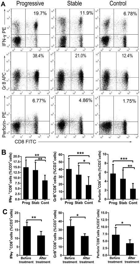
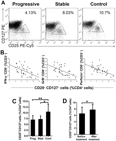
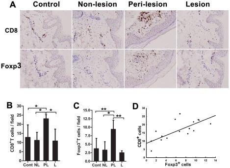
Melanocyte-specific CD8(+) cytotoxic T lymphocytes (CTLs) play a pivotal role in vitiligo-induced depigmentation. Yet, the mechanisms underlying the high frequency of generalized autoimmune disorders associated with generalized vitiligo (GV) are unknown. We hypothesized that an imbalance between activated CD8(+) CTLs and regulatory T cells (Tregs) exists in patients with GV . Assessment of the circulating CD8(+) CTLs and Tregs by flow cytometric analysis revealed an obvious expansion of CD8(+) CTLs and a concomitant decrease in Treg cells in GV patients. The percentages of skin infiltrating CD8(+) CTLs and Tregs were evaluated by immunohistochemistry and revealed dramatically increased numbers of both CD8(+) CTLs and Tregs in the perilesional skin of GV patients. However, peripheral Tregs were impaired in their ability to suppress the proliferation and cytolytic capacity of autologous CD8(+) T cells, suggesting that a functional failure of Tregs and the hyper-activation of CD8(+) CTLs may contribute to progressive GV. Our data indicate that reduced numbers and impaired function of natural Tregs fail to control the widespread activation of CD8(+) CTLs, which leads to the destruction of melanocytes and contributes to the elevated frequency of various associated autoimmune diseases. This knowledge furthers our understanding of the mechanisms of immune tolerance that are impaired in GV patients and may aid in the future development of effective immunotherapy for GV patients.


Conflict of interest statement

Competing Interests: The authors have declared that no competing interests exist.

Figures

Figure 1
Figure 1. Circulating CD8+ CTLs are increased significantly in GV patients.
(A) IFN-γ, GrB and Perforin–expressing cells were detected by intracellular staining. Values are the percentage of IFN-γ+ CD8+, GrB+ CD8+ or Perforin+ CD8+ cells among CD3+ T cells (Representative cases). (B) Flow cytometric analysis of IFN-γ+ CD8+, GrB+ CD8+ and Perforin+ CD8+ cells within the CD3+ T cell populations from patients with progressive GV (n = 38), stable GV (n = 12) and control subjects (n = 20). * = P<0.05, ** = P<0.01, *** = P<0.001. (C) The CD8+ CTL population of 6 patients was monitored longitudinally: The percentage of IFN-γ+ CD8+ T cells, GrB+ CD8+ T cells and Perforin+ CD8+ T cells was measured initially during GV progression and again following stabilization after treatment. * = P<0.01, ** = P<0.001.
Figure 2
Figure 2. Circulating CD4+ CD25+ CD127 Tregs are decreased significantly in GV patients.
(A) Human PBMCs were labeled with lymphocyte-specific antibodies (CD4, CD25 and CD127). Representative flow data showing the gating strategy and percent of natural Treg cells within the CD4+ T cell population are given for one patient from each of the 3 studied groups. (B) A negative correlation was found between the frequency of CD4+ natural Treg cells and the expression of IFN-γ (r = −0.512, P = 0.001), GrB (r = −0.524, P = 0.001) or Perforin (r = −0.585, P<0.001) from CD3+ CTLs in patients with progressive GV (n = 38). (C) The results of the flow cytometric analysis of natural Treg cells in patients with progressive GV (n = 38), stable GV (n = 12) and control subjects (n = 20) are shown. * = P<0.01, ** = P<0.001. (D) Longitudinal monitoring of natural Treg cells in 6 patients. The percentage of CD4+ natural Treg cells was measured twice: once during a vitiligo flare and once following resolution after treatment. * = P<0.05.
Figure 3
Figure 3. Skin-infiltrating CD8+ cells and Foxp3+ cells in GV patients.
(A) Consecutive sections were used for immunohistochemical detection of T cells expressing CD8 or Foxp3+ in skin samples from healthy controls or GV patients (representative fields, 200×). Positive cells appear brown. Both CD8+ (B) and Foxp3+ (C) T cell numbers were increased significantly in the perilesional (PL) tissues of progressive GV patients (n = 16). In contrast, no significant differences were observed between lesional (L) (n = 2) and non-lesional GV skin (NL) (n = 8), as compared to normal skin from unaffected adults (Cont) (n = 6). * = P<0.05, ** = P<0.01. (D) The number of CD8+ and Foxp3+ T cells in perilesional GV skin samples exhibited a positive correlation (r = 0.706, P<0.001; n = 16). Values in B–C are the mean and SD.
Figure 4
Figure 4. Tregs suppress proliferation and cytokine production of autologous CD8+ T cells.
(A) Detection of Treg suppression for CD8+ T cells. CFSE-labeled CD8+ T cells were stimulated in the presence of CD4+ CD25+ Treg cells at the indicated ratios for 5 days. Cellular division was measured by flow cytometric analysis based on the intensity of CFSE signal. Representative CFSE profiles from a GV patient and a normal control are shown. Values given represent the percentage of CFSElow cells among CFSE+ T cells. (B) Treg cells are largely unresponsive to TCR stimulation. Unlabeled and CFSE labeled CD4+ CD25+ T cells were cultured with anti-CD3/CD28 Abs, under the same stimulatory conditions as above, but without autologous CD8+ T cells present. Values given are the percentage of CFSElow cells among CFSE+ Treg cells (Representative cases). (C) Dose-dependent suppression of CD8+ T cell proliferation by CD4+ CD25+ T cells. The suppressive capacity of CD4+ CD25+ Tregs was compared between GV patients (n = 6) and normal control subjects (n = 4). * = P<0.05; ** = P<0.01. The production of IFN-γ (D) and TNF-α (E) was determined. CD8+ T cells were cultured with Tregs at various ratios in the presence of anti-CD3/CD28 Ab stimulation for 48 hours. IFN-γ and TNF-α release into the supernatant was determined by an enzyme-linked immunosorbent assay. The data were from 6 progressive GV individuals and 4 healthy controls. * = P<0.05; ** = P<0.01.

References

    1. Taieb A, Picardo M, Members V. The definition and assessment of vitiligo: a consensus report of the Vitiligo European Task Force. Pigment Cell Research. 2007;20:27–35. - PubMed
    1. Alkhateeb A, Fain PR, Thody A, Bennett DC, Spritz RA. Epidemiology of vitiligo and associated autoimmune diseases in Caucasian probands and their families. Pigment Cell Res. 2003;16:208–214. - PubMed
    1. Ogg GS, Dunbar PR, Romero P, Chen JL, Cerundolo V. High frequency of skin-homing melanocyte-specific cytotoxic T lymphocytes in autoimmune vitiligo. Journal of Experimental Medicine. 1998;188:1203–1208. - PMC - PubMed
    1. Palermo B, Campanelli R, Garbelli S, Mantovani S, Lantelme E, et al. Specific cytotoxic T lymphocyte responses against Melan-A/MART1, tyrosinase and Gp100 in vitiligo by the use of major histocompatibility complex/peptide tetramers: the role of cellular immunity in the etiopathogenesis of vitiligo. Journal of Investigative Dermatology. 2001;117:326–332. - PubMed
    1. Lang KS, Caroli CC, Muhm A, Wernet D, Moris A, et al. HLA-A2 restricted, melanocyte-specific CD8(+) T lymphocytes detected in vitiligo patients are related to disease activity and are predominantly directed against MelanA/MART1. Journal of Investigative Dermatology. 2001;116:891–897. - PubMed

Publication types

Substances