

Cell phenotypes in human amniotic fluid
- PMID: 22649611
- PMCID: PMC3347518
Cell phenotypes in human amniotic fluid
Abstract
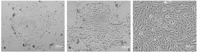
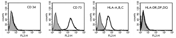
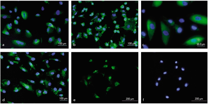
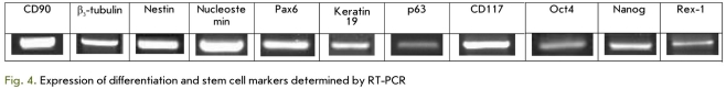
Stem cells capable of long-term proliferation and differentiation into different cell types may be a promising source of cells for regenerative medicine. Recently, much attention has been paid to fetal stem cells, among which are cells from amniotic fluid (AF). We have isolated amniotic stem cells from 3 AF samples. Flow cytometry, RT -PCR and immunohistochemistry have shown that these cells express mesenchymal (CD90, CD73, CD105, CD13, CD29, CD44, and CD146), neural (≤3-tubulin, Nestin, and Pax6), epithelial (keratin 19 and p63) markers and also markers of pluripotency (Oct4, Nanog, and Rex-1). Transplantation of the cells to nude mice does not lead to tumor formation. Thus, putative stem/progenitor cells from AF are capable of long-term proliferation in vitro and the profile of gene expression led us to speculate that they have greater differentiation potential than mesenchymal stem cells and may be useful for cell therapy.
Figures
References
-
- Baranov V.S., Kuznetsova T.V.. Cytogenetics of Human Embryonic Development. St. Petersburg. N-L. 2007:639–639.
-
- Bertani N., Matatesta P., Volpi G., Sonego P., Perris R. . Neurogenic Potential of Human Mesenchymal Stem Cells Revisited: Analysis by Immunostaining, Timelapse Video and Microarray. Journal of Cell Science. 2005;118:3925–3936. - PubMed
-
- Campagnoli C., Roberts I. A., Kumar S., Bennett P. R., Bellantuono I., Fisk N. M. . Identification of Mesenchymal Stem-Progenitor Cells in Human First-Trimester Fetal Blood, Liver and Bone Marrow. Blood. 2001;98:2396–2402. - PubMed
-
- Carraro G., Perin L., Sedrakyan S., Giuliani S., Tiozzo C., Lee J., Turcatel G., De Langhe S. P., Driscoll B., Bellusci S., Minoo P., Atala A., De Filippo R. E., Warburton D. . Human Amniotic Fluid Stem Cells Can Integrate and Differentiate into Epithelial Lung Lineages. Stem Cells. 2008;26:2902–2911. - PMC - PubMed
-
- Corti S., Locatelli F., Strazzer S., Guglieri M., Corni G. P. . Neuronal Generation from Somatic Stem Cells: Current Knowledge and Perspectives on the Treatment of Acquired and Degenerative Central Nervous System Disorders. Curr. Gene Ther. 2003;3:247–272. - PubMed
LinkOut - more resources
Full Text Sources
Other Literature Sources
Research Materials
Miscellaneous