. 2009 Oct;1(3):29-51.

Chemical and functional aspects of posttranslational modification of proteins

Affiliations

Chemical and functional aspects of posttranslational modification of proteins

D G Knorre et al. Acta Naturae. 2009 Oct.

Abstract

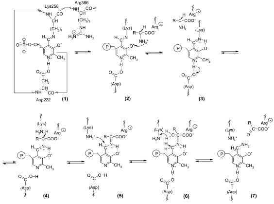
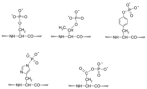
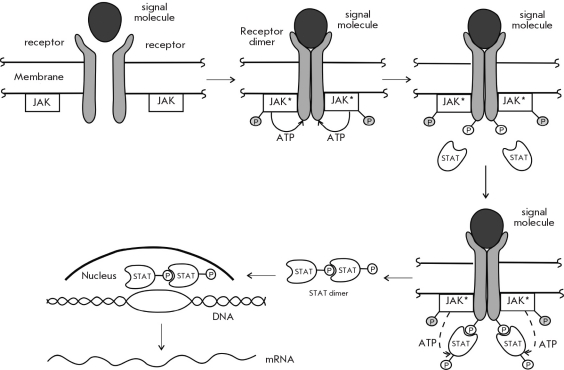
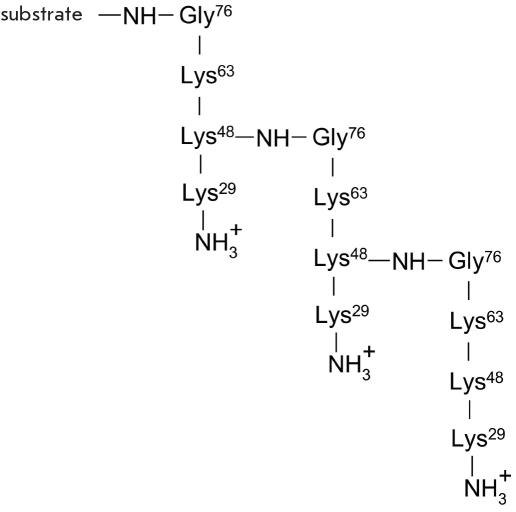
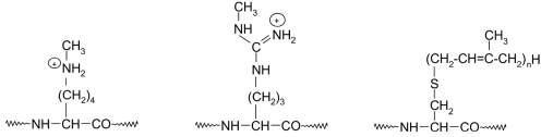
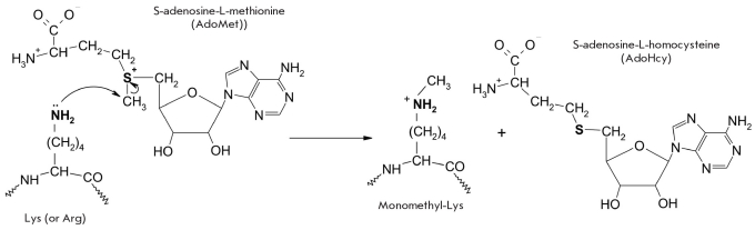
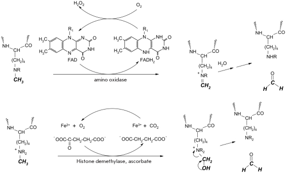
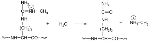
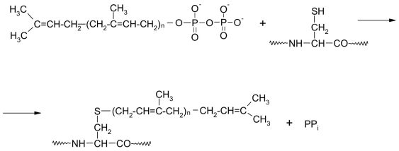
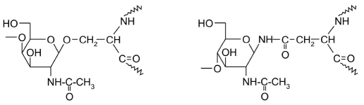
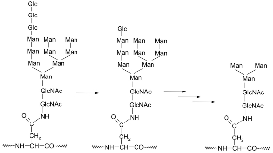
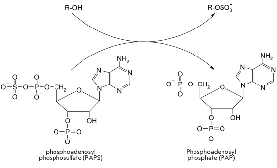
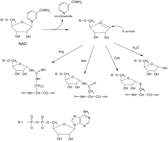
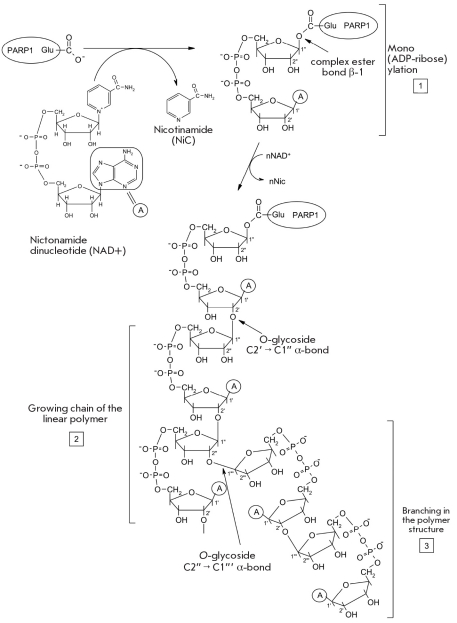
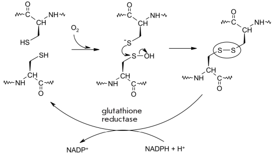
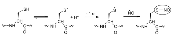
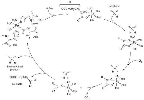
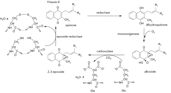
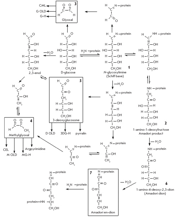
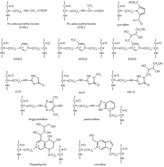
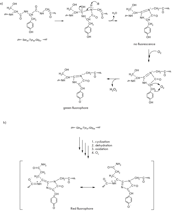
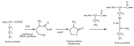
This paper reviews the chemical and functional aspects of the posttranslational modifications of proteins, which are achieved by the addition of various groups to the side chain of the amino acid residue backbone of proteins. It describes the main prosthetic groups and the interaction of these groups and the apoenzyme in the process of catalysis, using pyridoxal catalysis as an example. Much attention is paid to the role of posttranslational modification of proteins in the regulation of biochemical processes in live organisms, and especially to the role of protein kinases and their respective phosphotases. Methylation and acetylation reactions and their role in the "histone code", which regulates genome expression on the transcription level, are also reviewed. This paper also describes the modification of proteins by large hydrophobic residues and their role in the function of membrane-associated proteins. Much attention is paid to the glycosylation of proteins, which leads to the formation of glycoproteins. We also describe the main non-enzymatic protein modifications such as glycation, homocysteination, and desamida-tion of amide residues in dibasic acids.


Figures

Fig. 1.
Fig. 1.
A schematic representation of the first stage of the transamination reaction catalyzed by aspartate aminotransferase
Fig. 2.
Fig. 2.
The structure of phosphorylated amino acid fragments
Fig. 3.
Fig. 3.
The basic stages of signal transduction via protein phosphorylation. IF - inositoltriphosphate, DAG - diacylglycerine
Fig. 4.
Fig. 4.
A schematic representation of signal transduction via JAK-associated membrane receptors
Fig. 5.
Fig. 5.
Addition of an ubiquitin residue (residues) onto a substrate protein. E1-SH - ubiquitin-activating enzyme, E2-SH - ubiquitin transport protein, E3 - ubiquitin-protein ligase. Ub - ubiquitin residue
Fig. 6.
Fig. 6.
A tandem of several ubiquitin residues bound to the substrate. The numbers refer to the amino acid residues which take part in the modification of the substrate (Gly76) and formation of the tandem (Gly76 and Lys48)
Fig. 7.
Fig. 7.
Structures of alkylated amino acid side chains in proteins
Fig. 8.
Fig. 8.
Methylation of lysine residues by methyltransferases
Fig. 9.
Fig. 9.
Demethylation reaction of di- and monomethylated lysine residues in histones catalyzed by the FAD-dependent aminooxidase (top), and tri-, di- and monomethylated lysine residues in histones catalyzed by histone demethylase, which functions in the presence of cofactors, Fe2+ ions, Alpha;-ketoglutarate and ascorbate (bottom)
Fig. 10.
Fig. 10.
Demethylation of modified arginine residues catalyzed by the nuclear peptidylarginine deiminase (PAD4) [58]
Fig. 11.
Fig. 11.
Tranfer of an isoprenoid residue from pyrophosphate to a cysteine residue in an apoprotein. n = 2 - is a farnseyl residue, n = 3 - geranyl-geranyl residue
Fig. 12.
Fig. 12.
Prenylation of the Ras protein: 1 - addition of a farnesyl residue onto the Cys-A-A-X sequence (A- a small aliphatic amino acid residue, X is Leu, Phe or Met); 2 - Cleaving of the A-A-X tripeptide by the Rasconverting enzyme, which is a CysAAX-endopeptidase; 3 - carboxymethylation of the isoprenylcysteine residue catalyzed by the isoprenylcysteine carboxymethyltransferase [86]
Fig. 13.
Fig. 13.
Structures of the products of N-acetylglucosamine addition onto serine and asparagine side chains in proteins
Fig. 14.
Fig. 14.
Structure of the carbohydratebearing dolichol pyrophosphate
Fig. 15.
Fig. 15.
Structure and first stages of the processing of oligosaccharide fragment Glc3Man9(GlcNAc)2 as a part of a glycoprotein. Monosaccharides Glc - glucose, GlcNAc - N-acetylglucosamine
Fig. 16.
Fig. 16.
Sulfation reaction catalyzed by sulfotransferase
Fig. 17.
Fig. 17.
(ADPribosyl) ation of nucleophilic amino acid residues (X) present in the protein (cysteine, arginine and asparagine) [1]
Fig. 18.
Fig. 18.
Modification of the His715 residue in the structure of the human eEF-2 elongation factor results in the blocking of protein synthesis in human cells
Fig. 19.
Fig. 19.
Mechanism of poly(ADP-ribose) synthesis in the self-modification of PARP1
Fig. 20.
Fig. 20.
Oxidation of the sulfhydryl group of the cysteine residue, which can he reduced again into a thiol group with the help of NAD(P) H and a glutathione reductase [147]
Fig. 21.
Fig. 21.
Oxidation of the thiolate ion in the presence of nitrogen oxide results in the formation of cysteinyl-nitroxide [154]
Fig. 22.
Fig. 22.
Structure of monooxygenated proline, lysine, and asparagine residues
Fig. 23.
Fig. 23.
Mechanism of the hydroxylation reaction
Fig. 24.
Fig. 24.
Vitamin K-dependent carboxylation of a glutamic acid residue catalyzed by γ-glutamylcarboxylase. The 2,3-epoxide of vitamin K is reduced by vitamin K 2,3-epoxide reductase
Fig. 25.
Fig. 25.
Glycation of proteins in the presence of D-glucose. The rectangles show the main precursors of AGEs, which are formed during glycation
Fig. 26.
Fig. 26.
Structure of certain AGEs formed as a result of in vivo protein modification by D glucose
Fig. 27.
Fig. 27.
Formation of (a) green and (b) red chromophores in proteins from tripeptides by intramolecular posttranslational autocatalytic cyclization
Fig. 28.
Fig. 28.
N-homocysteinylation of proteins by the homocysteine thiolactone
Fig. 29.
Fig. 29.
Protein S-homocysteinylation
Fig. 30.
Fig. 30.
Deamidation of asparagine residues in peptides and proteins at pH>5
Fig. 31.
Fig. 31.
Transamidiation catalyzed by transglutaminase (E.C. 2.3.2.13)

References

    1. Walsh C.T., Garneau-Tsodikova S., Gatto G.J. Angew. Chem. Int. Ed. 2005;44(45):7342–7372. - PubMed
    1. Lehninger A. Principles of Biochemistry. New York: W.H. Freeman and Company; 2008.
    1. Karpeysky M.Y., Ivanov V.I. Nature. 1966;210(30):493–496. - PubMed
    1. Lowe J.N., Ingraham L.L. An Introduction to Biochemical Reactions Mechanisms. New Jersey: Prentice-Hall: Englewood Cliffs; 1974. Chap. 3. Foundation of Molecular Biology Series.
    1. Hubbard S.R. Handbook of Cell Signaling. 2009. Chap. 58; pp. 413–418.

LinkOut - more resources