. 2010 Apr;2(1):72-82.

Calcium signaling and neurodegeneration

Affiliations

Calcium signaling and neurodegeneration

I B Bezprozvanny. Acta Naturae. 2010 Apr.

Abstract

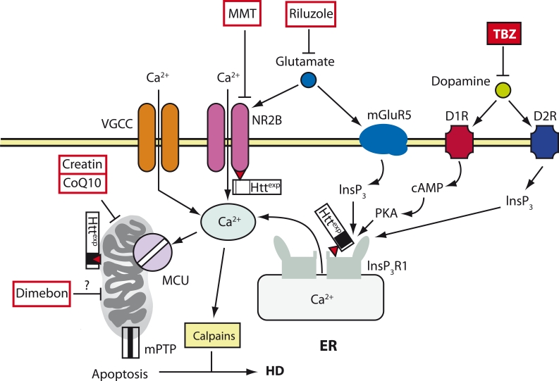
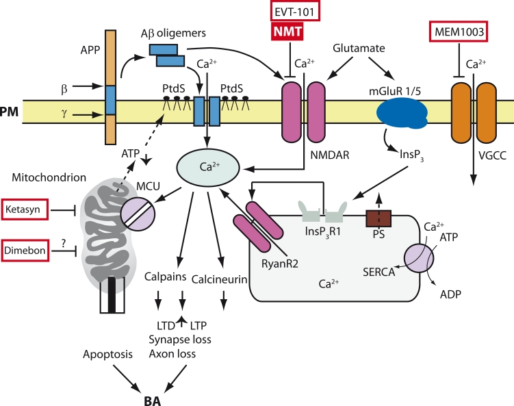
Neurodegenerative disorders, such as Alzheimer's disease (AD), Parkinson's disease (PD), amyotrophic lateral sclerosis (ALS), Huntington's disease (HD), and spinocerebellar ataxias (SCA) are very important both for fundamental science and for practical medicine. Despite extensive research into the causes of these diseases, clinical researchers have had very limited progress and, as of now, there is still no cure for any of these diseases. One of the main obstacles in the way of creating treatments for these disorders is the fact that their etiology and pathophysiology still remain unclear. This paper reviews results that support the so-called "calcium hypothesis of neurodegenerative diseases." The calcium hypothesis states that the atrophic and degenerative processes in the neurons of AD, PD, ALS, HD, and SCA patients are accompanied by alterations in calcium homeostasis. Moreover, the calcium hypothesis states that this deregulation of calcium signaling is one of the early-stage and key processes in the pathogenesis of these diseases. Based on the results we reviewed, we conclude that the calcium channels and other proteins involved in the neuronal calcium signaling system are potential drug targets for AD, PD, ALS, HD, and SCA therapy.

Keywords: Alzheimer’s disease; Huntington’s disease; Parkinson’s disease (PD); amyotrophic lateral sclerosis; calcium channels; calcium signaling; clinical trials; dimebon; imaging; memantine; mitochondria; riluzole; spinocerbellar ataxias; transgenic mice.


Figures

Fig. 1
Fig. 1
A model of Ca2+ deregulation during HD (cited from (Tang et al., 2007)). In MSNs during HD, Httexp disrupts Ca2+ signaling by three syn- ergistic mechanisms. Httexp increases the function of the NR2B-bearing NMDA receptor (probably by increasing its transport into the plasma membrane). Httexp tightly binds to the С-terminus of InsP3R1 and increases its affinity to InsP3. The low level of glutamate secreted by the neurons of the corticostriatal projection causes an excessive influx of Ca2+ via the NMDA receptor and the release of Ca2+ from the ER via InsP3R1. The additional uptake of Ca2+ into MSNs is mediated by VGCC. Dopamine excreted by the dopaminergic neurons of the mesencephalon stimulates the type-1 (D1R) and type-2 (D2R) dopamine receptors, which are highly expressed in MSN. D1R is connected with an adenylate cyclase, and together they increase the cAMP level and activate the protein kinase A (PKA). PKA enhances the glutamate-induced Ca2+ signals by increasing the activity of the NMDA receptor and InsP3R1. D2R is directly involved in the produc- tion of InsP3 and the activation of InsP3R1. The excessive uptake of Ca2+ activates calpain, which cleaves Httexp and other substrates. The excess of Ca2+ in the cytosol leads to the capture of Ca2+ by the mitochondria via MCU, which in turn induces the opening of mPTP and apoptosis. The calcium regulation of mitochondria is also disrupted due to the direct interaction between Httexp and the mitochondria. The antidopamine drug tetrabenzine (TBZ) has been approved in the United States for the symptomatic treatment of HD. The NMDA receptor antagonist memantin (MMT), the soluble “mitochondrial agent” dimebon and “mitochondrial stabilizers” creatin and coenzyme Q10 (CoQ10) are all in clinical trials. The antiglutamate agent Riluzole has passed clinical trials, but it turned out to be ineffective for HD treatment [19]
Fig. 2
Fig. 2
Model of Ca2+ deregulation in AD (Bezprozvanny and Mattson, 2008). Sequential cleavage of the β-amyloid precursor-protein (β) by the γ-secretase (γ) leads to the formation of Aβ. Aβ forms oligomers, which can integrate into the plasma membrane (PM) and form pores permeable to Ca2+-ions. The association of Aβ oligomers with the plasma membrane is facilitated by binding with the surface phosphatidylserine (PtdS); the aging process and Ca2+-mediated damage of mitochondria cause a decrease in the ATP level and lead to the transfer of PtdS from the inner to the outer surface of the plasma membrane. The decrease of the ATP level and the loss of membrane integrity cause membrane depolarization, which in turn causes an increase in the uptake of Ca2+ through NMDA receptors and VGCC. Aβ oligomers can also directly influence the affinity of the NMDA, AMPA, and VGCC receptors. Gluta- mate activates mGluR1/5 receptors, increases the production of InsP3, and facilitates InsP3-mediated release of Ca2+ from the ER. Presenilins (PS) function as channels for Ca2+ drain from the ER and various muta- tions associated with HAD disrupt the Ca2+ drain function of presenilins. This causes the excessive accumulation of Ca2+ in the ER. An increase in Ca2+ levels in the ER enhances the release of Ca2+ via the type-I InsP3 (InsP3R1) and type-2 ryanodine receptor (RyanR2). PS can also directly modulate the activity of InsP3R, RyanR, and the SERCA pump. The increase in cytosolic Ca2+ concentration activates calcineurin and calpains, which in turn enhances long-term depression (LTD), suppresses long-term potentiation (LTP), and causes the modification of the neuronal cytoskeleton and the loss of synapses and axon atrophy. The excessive amount of Ca2+ in the mitochondria, which appears due to the activity of mitochondrial calcium uniporter (MCU), leads to the opening of the mitochondrial permeable transit pore (mPTP) and apoptosis. An inhibitor of the NMDA receptor called memantine (MMT) has been approved for AD treatment, and a NR2B-specific antagonist EVT-101 has also been developed. Currently, several other drugs are under clinical trials for the treatment of AD: a “CNS optimized” L-type VGCC inhibitor MEM-1003, the soluble “mitochondrial agent” Dimebon, and “mitochondrial antide- pressant ” Ketasyn

References

    1. Berridge M.J.. Neuronal Calcium Signaling. Neuron. 1998;21:13–26. - PubMed
    1. Toescu E.C, Verkhratsky A. . The Importance of Being Subtle: Small Changes in Calcium Homeostasis Control Cognitive Decline in Normal Aging. Aging Cell. 2007;6:267–273. - PubMed
    1. Gant J.C., Sama M.M., Landfield P.W, Thibault O.. Early and Simultaneous Emergence of Multiple Hippocampal Biomarkers of Aging is Mediated by Ca2+-Induced Ca2+ . 2006;26:3482–3490. - PMC - PubMed
    1. Foster T.C.. Calcium Homeostasis and Modulation of Synaptic Plasticity in the Aged Brain. Aging Cell. 2007;6:319–325. - PubMed
    1. Gusella J.F., MacDonald M.E.. Molecular Genetics: Unmasking Polyglutamine Triggers in Neurodegenerative Disease. Nat Rev Neurosci. 2000;1:109–115. - PubMed

LinkOut - more resources