. 2010 Jul;2(2):102-6.

Derivation of induced pluripotent stem cells from fetal human skin fibroblasts

Affiliations

Derivation of induced pluripotent stem cells from fetal human skin fibroblasts

S P Medvedev et al. Acta Naturae. 2010 Jul.

Abstract

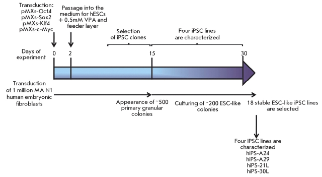
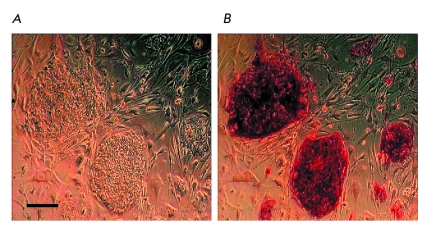
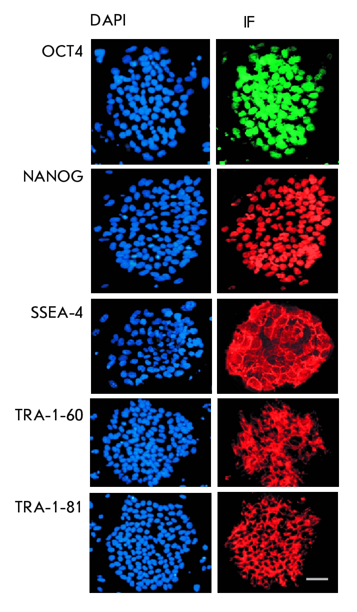
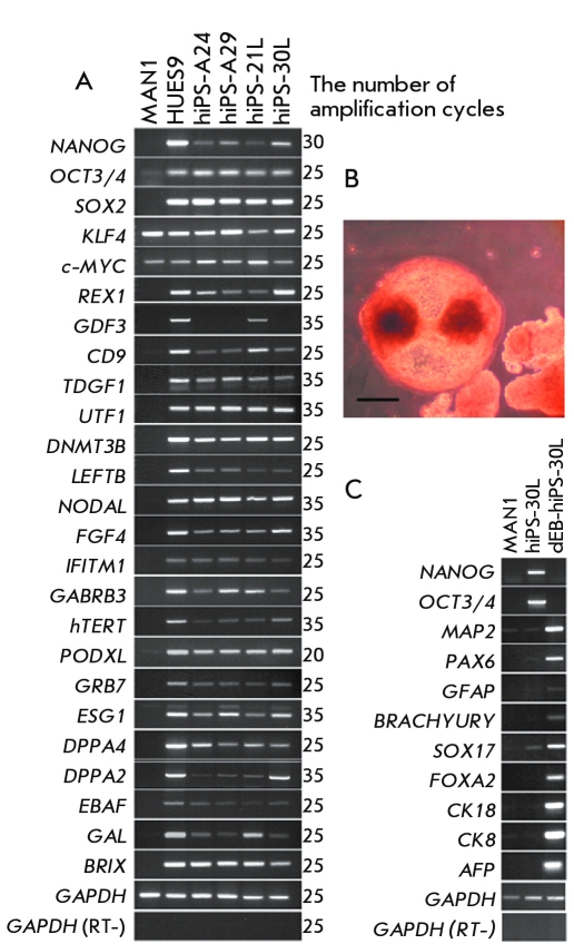
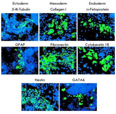
The isolation and study of autologous human stem cells remain among the most urgent problems in cell biology and biomedicine to date. Induced pluripotent stem cells can be derived from human somatic cells by the overexpression of a number of genes. In this study we reprogrammed fetal human skin fibroblasts by transduction with retroviral vectors carrying murine Oct4 , Sox2 , Klf4 , and c-Myc cDNAs. As a result, cells with the protein expression and gene transcription pattern characteristic of human embryonic stem cells were derived. These induced pluripotent cells are capable of differentiation in vitro into the ectoderm, mesoderm, and endoderm derivatives.

Keywords: induced pluripotent stem cells; reprogramming; retroviral vectors.


Figures

Fig. 1
Fig. 1
Schematic representation of an experiment on the preparation of induced pluripotent stem cells from human embryonic fibroblasts.
Fig. 2
Fig. 2
(A) morphology of iPSC colonies derived from hEFs; (B) staining of the iPSC colonies demonstrating the expression of alkaline phosphatase (one of the pluripotent cell markers). Scale 100 µm.
Fig. 3
Fig. 3
Immunocytochemical staining (IF) of iPSC colonies with antibodies against the transcription factors OCT4 (green) and NANOG (red) and against the surface antigens SSEA-4, TRA-1-60, and TRA-1-81 (red). Cell nuclei are counterstained with DAPI (blue). Scale 100 µm.
Fig. 4
Fig. 4
(A) RT-PCR assay of gene transcripts, which are the markers of human embryonic stem cells, in embryonic fibroblasts (MA N1), human embryonic stem cells (HUES9), and four iPSC lines: hiPS-A24, hiPS-A29, hiPS-21L, and hiPS-30L; (B) embryoid bodies formed following the passage of hiPS-30l iPSC cells into a liquid medium, scale 100 µm; (C) RT-PCR assay of gene transcripts, which are the markers of three germ layers (ectoderm, mesoderm, and endoderm), following the differentiation of the hiPS-30L clone via the embryoid body formation.
Fig. 5
Fig. 5
Expression of gene markers characterizing three germ layers (ectoderm, mesoderm, and endoderm) in spontaneous iPSC differentiation in embryoid bodies derived from hEFs. Scale 100 µm.

References

    1. Yamanaka S.. Cell. 2009;137:13–17. - PubMed
    1. Park I.H., Arora N., Huo H.. Cell. 2008;134:877–886. - PMC - PubMed
    1. Ebert A.D., Yu J., Rose F.F.. Nature. 2009;457:277–280. - PMC - PubMed
    1. Lee G., Papapetrou E.P., Kim H.. Nature. 2009;461:402–406. - PMC - PubMed
    1. Takahashi K., Yamanaka S.. Cell. 2006;126:663–676. - PubMed

LinkOut - more resources