. 2011 Jan;3(1):48-68.

Assays for detection of telomerase activity

Affiliations

Assays for detection of telomerase activity

D A Skvortsov et al. Acta Naturae. 2011 Jan.

Abstract

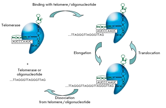
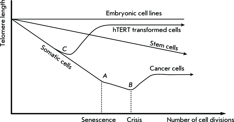
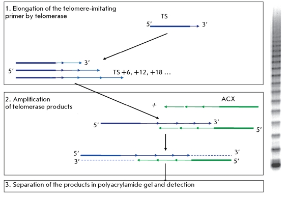
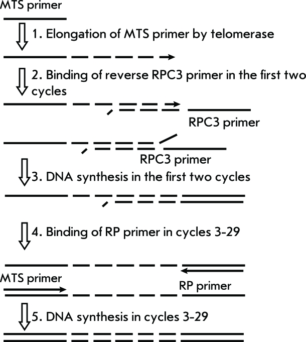
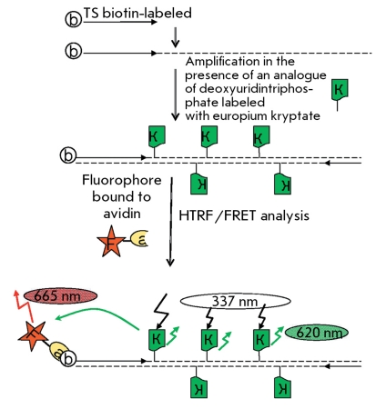
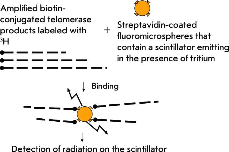
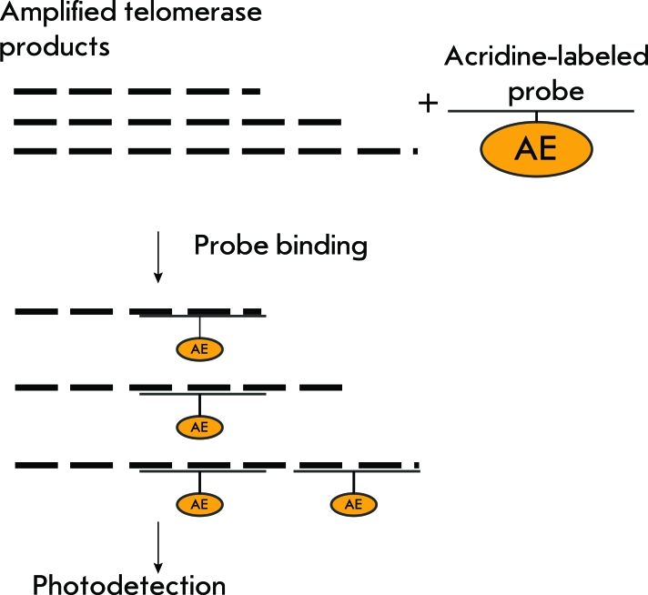
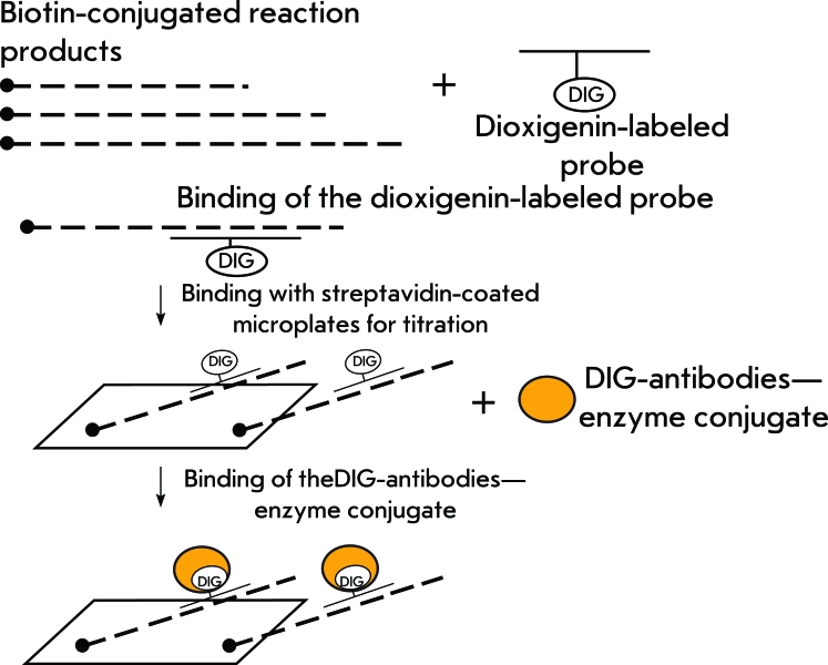
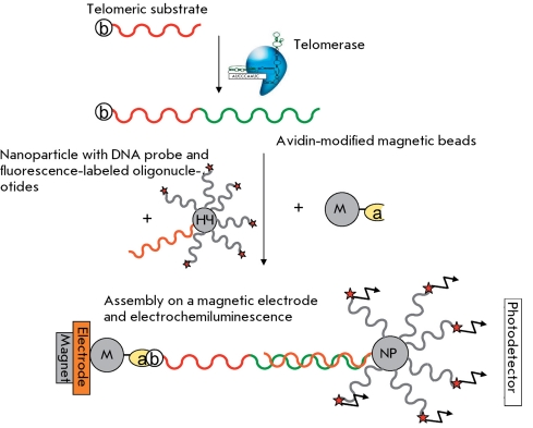
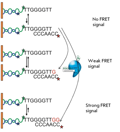
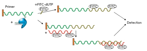
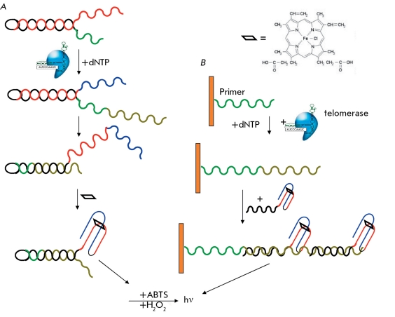
Progressive loss of the telomeric ends of chromosomes caused by the semi-conservative mechanism of DNA replication is an important timing mechanism which controls the number of cells doubling. Telomerase is an enzyme which elongates one chain of the telomeric DNA and compensates for its shortening during replication. Therefore, telomerase activity serves as a proliferation marker. Telomerase activity is not detected in most somatic cells, with the exception of embryonic tissues, stem cells, and reproductive organs. In most tumor cells (80-90%), telomerase is activated and plays the role of the main instrument that supports the telomere length, which can be used for the diagnostics of neoplastic transformation. This is the primary reason why assays regarding the development of telomerase activity have attracted the attention of researchers. Telomerase activity testing may be useful in the search for telomerase inhibitors, which have the potential to be anti-cancer drugs. Moreover, telomerase activation may play a positive role in tissue regeneration; e.g., after partial removal of the liver or cardiac infarction. All telomerase activity detection assays can be divided into two large groups: those based on direct detection of telomerase products, and those based on different systems of amplification of the signals from DNA that yield from telomerase. The methods discussed in this review are suitable for testing telomerase activity in different samples: in protozoa and mammalian cells, mixed cellular populations, and tissues.

Keywords: DNA determination; physicochemical methods; polymerase activity assay; telomerase; telomerase activity assay; tumor diagnostics.


Figures

Fig. 1
Fig. 1
General functional scheme of the telomerase complex.
Fig. 2
Fig. 2
Dependence of telomere length on the number of cell divisions in different cell types: embryonic cell lines, somatic cells, hTERT-transformed cells. A– Hayflick limit, B – crisis with subsequent apoptosis or tumor transformation, C – cell transformation by hTERT gene.
Fig. 3
Fig. 3
Telomeric repeat amplification protocol (TRAP).
Fig. 4
Fig. 4
“Two-primer” TRAP scheme [23, 24].
Fig. 5
Fig. 5
Scheme of the TRAP method with time-resolution fluorescence energy resonance transfer [26]. F – fluorophore, a – avidin, b –biotin, and K – analog of deoxyuridintriphosphate labeled with europium kryptate.
Fig. 6
Fig. 6
Scheme for TRAP with scintillation proximity assay.
Fig. 7
Fig. 7
Scheme for TRAP with hybridization protection assay.
Fig. 8
Fig. 8
Scheme for ELISA detection of PCR products in TRAP-ELISA (or detection of telomerase reaction products in direct ELISA).
Fig. 9
Fig. 9
Scheme of telomerase activity detection with the PCR amplification of telomerase products for transcriptional amplification [12, 42].
Fig. 10
Fig. 10
Scheme of the use of surface plasmon resonance (SPR) for detecting macromolecules; (A) sensogram corresponding to the general scheme and (B) SPR sensogram for telomerase activity detection. RU – resonance units. The difference between signals 1 and 2 represents DNA that was synthesized by telomerase [44].
Fig. 11
Fig. 11
Telomerase magnetic nanosensor. A - Complex assembly between the synthesized telomere repeats and oligonucleotide magnetic nanoparticles conjugated with antitelomere. B - Induced magnetic T2 changes as a function of time after adding an oligonucleotide consisting of either a 54-bp telomeric repeat or the primer [46].
Fig. 12
Fig. 12
Processes on the sensor in quartz crystal microbalance and electrochemical methods. A - Insertion of a biotin-labeled dUTP into the telomerase-synthesized DNA. B - Hybridization of biotin-labeled oligonucleotide with telomerase-synthesized DNA [47]. b – biotin as a component of the oligonucleotide, a-AP –avidin–alkaline phosphatase conjugate, dUTP – mixture of nucleoside triphosphates, biot-dUTP – biotin-labeled dUTP.
Fig. 13
Fig. 13
Assembly on the electrochemically detectable complex upon telomerase activity detection with ferrocenylnaphthalene diimide [48].
Fig. 14
Fig. 14
Biobarcode-based electrochemical telomerase detection [49]. NP – nanoparticles.
Fig. 15
Fig. 15
A - The scheme of formation of telomerase-synthesized DNA on the sensor surface. B - Sensogram for determination of the surface loading on the optical sensor [50].
Fig. 16
Fig. 16
A - Telomerization of the oligonucleotide bound with CdSe-ZnS by a quantum dot with the incorporation of Texas Red-labeled dUTP. B - Switching from the wavelength of quantum dot fluorescence (λ1) to Texas Red fluorescence (λ2) upon telomerization of the nucleotide bound with CdSe-ZnS by a quantum dot with the incorporation of TR-dUTP [51]. TR – Texas Red.
Fig. 17
Fig. 17
Electrochemiluminescence method for telomerase activity detection [54].
Fig. 18
Fig. 18
Telomerase activity detection using FRET and total internal reflection fluorescence [57].
Fig. 19
Fig. 19
Scheme of formation of fluorescence-labeled telomerase-synthesized DNA detection by a fluorimetric optical sensor [59].
Fig. 20
Fig. 20
(A, B) Two schemes for telomerase activity detection with DNAenzymes [60, 61].

References

    1. Hayflick L., Moorhead P.S.. Exp. Cell Res. 1961;25:585–621. - PubMed
    1. Olovnikov A.M.. J. Theor. Biol. 1973;41(1):181–190. - PubMed
    1. Blackburn E.H.. Cell. 2001;106(6):661–673. - PubMed
    1. Pandita T.K., Hunt C.R., Sharma G.G., Yang Q.. Cell Mol. Life Sci. 2007;64(2):131–138. - PMC - PubMed
    1. Greider C.W., Blackburn E.H.. Cell. 1985;43(2):405–413. - PubMed

LinkOut - more resources