. 2011 Jan;3(1):107-15.

Modeling myocardial infarction in mice: methodology, monitoring, pathomorphology

Affiliations

Modeling myocardial infarction in mice: methodology, monitoring, pathomorphology

A A Ovsepyan et al. Acta Naturae. 2011 Jan.

Abstract

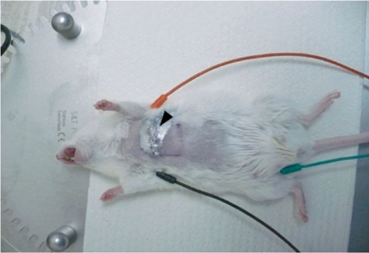
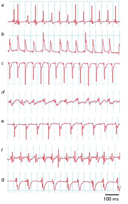
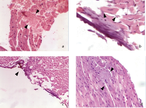
Myocardial infarction is one of the most serious and widespread diseases in the world. In this work, a minimally invasive method for simulating myocardial infarction in mice is described in the Russian Federation for the very first time; the procedure is carried out by ligation of the coronary heart artery or by controlled electrocoagulation. As a part of the methodology, a series of anesthetic, microsurgical and revival protocols are designed, owing to which a decrease in the postoperational mortality from the initial 94.6 to 13.6% is achieved. ECG confirms the development of large-focal or surface myocardial infarction. Postmortal histological examination confirms the presence of necrosis foci in the heart muscles of 87.5% of animals. Altogether, the medical data allow us to conclude that an adequate mouse model for myocardial infarction was generated. A further study is focused on the standardization of the experimental procedure and the use of genetically modified mouse strains, with the purpose of finding the most efficient therapeutic approaches for this disease.

Keywords: ECG (electrocardiogram); controlled electrocoagulation; coronary artery; ligation; myocardial infarction.


Figures

Fig. 1
Fig. 1
Preoperation and operation procedures during simulation of myocardial infarction in mice: a – fixation of a mouse on a heated operation table; b – intubation of trachea; c – device for artificial pulmonary ventilation “MiniVent Ventilator for Mice”; d – incision in the 4-th left intercostal space; e – front (lower) surface of the heart can be seen in the incision wound; f – ligation of the descending branch of the coronary artery.
Fig. 2
Fig. 2
Illustration of postsurgery ECG measurements. A mouse is placed on a heated operation table and connected to the ECG apparatus by three electrodes: one on the left foreleg (red), one on the right foreleg (black), and one on the right hind leg (black). The sutured wound is covered with an AluSpray suspension (shown with an arrow).
Fig. 3
Fig. 3
ECG recorded in animals after surgery: a – ECG recorded prior to surgery; b – impaired heart rhythm in 5–10 minutes after ligation of the coronary artery; c – development of a large-focal infarction 10 days after the ligation of the coronary artery; d – heart rhythm measured 5 minutes after thermal coagulation of the coronary artery; e – development of surface infarction 10 days after thermal coagulation; f – tachycardia developed 5–10 minutes after the ligation of the coronary artery; g – brachycardia developed on day 3 after the ligation of the coronary artery. The excitation and repolarization width is 100 ms.
Fig. 4
Fig. 4
Histopathology of the hearts in the case of infarction simulated in mice: a – beginning of coagulational necrosis and cardiomyocytes disorganization, stromal edema, and haemorrhage; hematoxylin and eosin staining; magnification of 20x; b – lack of cross striation within the sarcoplasm of cardiomyocytes; apoptotic patterns; hematoxylin and eosin staining; magnification of 60x; c – necrosis stripe of the epicardium and surface cardiomyocytes occurring under the impact of the electrode during coagulation; edema of the myocardial interstitium; hematoxylin and eosin staining; magnification of 10x; d – fragmentation of muscle fibers as a result of fibrillation; hematoxylin and eosin staining; magnification of 40x.

References

    1. Burgueño A.L., Landa M.S., Schuman M.L., Alvarez A.L., Carabelli J., García S.I., Pirola C.J.. Metabolism. 2007;56(10):1439–1443. - PubMed
    1. Cascio W.E., Cozzi E., Hazarika S., Devlin R.B., Henriksen R.A., Lust R.M., van Scott M.R., Wingard C.J.. Inhal Toxicol. 2007;19(5):67–73. - PubMed
    1. Cohen J.K., Cai L.Q., Zhu Y.S., La Perle K.M.. Comp. Med. 2007;57(4):370–376. - PubMed
    1. Denvir M.A., Sharif I., Anderson T., Webb D.J., Gray G.A., McDicken W.N.. J. Am. Soc. Echocardiogr. 2005;18(2):155–162. - PubMed
    1. Lee S., Schwinger R.H., Brixius K.. Pflügers Arch. 2008;455(5):767–774. - PubMed

LinkOut - more resources