. 2011 Oct;3(4):12-29.

Escort aptamers: new tools for the targeted delivery of therapeutics into cells

Affiliations

Escort aptamers: new tools for the targeted delivery of therapeutics into cells

A S Davydova et al. Acta Naturae. 2011 Oct.

Abstract

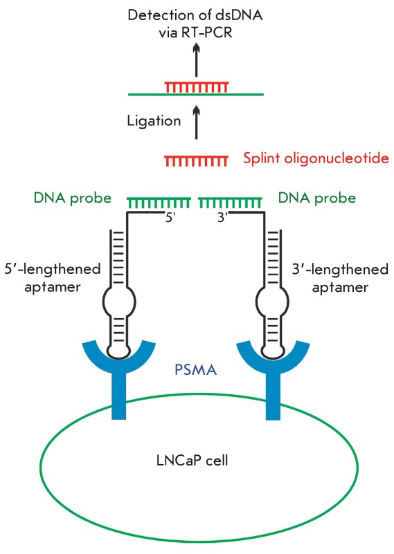
Escort aptamers are DNA or RNA sequences with high affinity to certain cell-surface proteins, which can be used for targeted delivery of various agents into cells of a definite type. The peculiarities of the selection of escort aptamers are discussed in this review. The methods used in selection of escort aptamers via the SELEX technique are considered, including selection against isolated cell-surface proteins, cell fragments, living eukaryotic cells, and bacteria. Particular attention is given to the design and chemical modification of escort aptamers. The different fields of application of escort aptamers are described, including the targeted delivery of siRNAs, nanoparticles, toxins, and photoagents, as well as the identification of specific cell markers and the detection or isolation of cells of a definite type. The potential for the application of escort aptamers in the development of new therapeutic agents and diagnostic systems is also discussed.

Keywords: NA aptamers; SELEX method; addressed cell delivery; detection of cells; escort aptamers; specific cell binding.


Figures

Fig. 1
Fig. 1
The general scheme of the SELEX method using DNA (A) and RNA (B) libraries.
Fig. 2
Fig. 2
Chemical modifications of sugar-phosphate backbone increasing the resistance of escort aptamers in biological media.
Fig. 3
Fig. 3
. The scheme of in vitro cell selection (including the negative selection stage) by the example of the RNA library.
Fig. 4
Fig. 4
The proposed secondary structure of the modified ТТА1 aptamer binding to tenascin C [19]. Designations: N F  – 2’-fluoro-2’-deoxyribonucleotide, N m  – 2’-O-methylribonucleotide, NH 2  – aminohexanol residue, L – hexaethylene glycol phosphate linker.
Fig. 5
Fig. 5
The proposed secondary structure of the modified C8FL aptamer against CCRF-CEM cells [18]. Designations: L – hexaethylene glycol phosphate, N *  – LNA nucleotides.
Fig. 6
Fig. 6
The proposed secondary structure of the trimer formed by the modified TD05.17 aptamer against Ramos cells [33]. Designations: L – hexaethylene glycol phosphate, N *  – LNA nucleotides.
Fig. 7
Fig. 7
The proposed secondary structure of DNA aptamer III.1 against microvessels of rat brain tumor [37].
Fig. 8
Fig. 8
The proposed secondary structure of RNA aptamer А07 against the human transforming growth factor receptor [44]. Designations: N F  – 2’-fluoro-2’-deoxyribonucleotide.
Fig. 9
Fig. 9
The proposed secondary structure of DNA aptamer ZE2 against the hepatitis C virus coat protein [70].
Fig. 10
Fig. 10
The main types of conjugates of escort aptamers used for targeted cell delivery and specific cell detection.
Fig. 11
Fig. 11
Schematic representation of chimeric constructs for siRNA delivery into PSMA-positive cells. A. The conjugate of biotinylated anti-PSMA aptamer and siRNA connected via streptavidin [74]. B. Chimeric RNA built from an anti-PSMA aptamer and siRNA [75]. Bio – biotin residue.
Fig. 12
Fig. 12
Detection of prostate-specific membrane antigen using anti-PSMA aptamers by the proximity ligation assay [96]. The method includes the following stages: 1) formation of the complementary complexes of aptamers with DNA probes; 2) binding of aptamers to the adjacent sites on the cell surface, thus positioning DNA probes closer to each other; 3) hybridization of both DNA probes with the splint oligonucleotide; 4) ligation of DNA probes in the resulting complex; 5) detection of dsDNA via real-time PCR.

References

    1. Ellington A.D., Szostak J.W.. Nature. 1990;346:818–822. - PubMed
    1. Tuerk C., Gold L.. Science. 1990;249:505–510. - PubMed
    1. Robertson D.L., Joyce G.F.. Nature. 1990;344:467–468. - PubMed
    1. Mayer G.. Angew. Chem. Int. Ed. 2009;48:2672–2689. - PubMed
    1. Stoltenburg R., Reinemann C., Strehlitz B.. Biomol. Eng. 2007;24:381–403. - PubMed

LinkOut - more resources