

Acute eosinophilic ascites in a middle-aged man
- PMID: 22649743
- PMCID: PMC3356866
- DOI: 10.1155/2012/896523
Acute eosinophilic ascites in a middle-aged man
Abstract
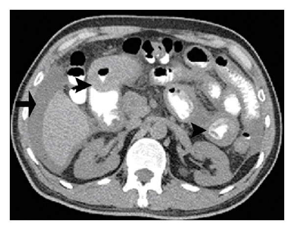
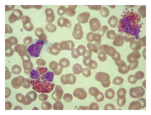
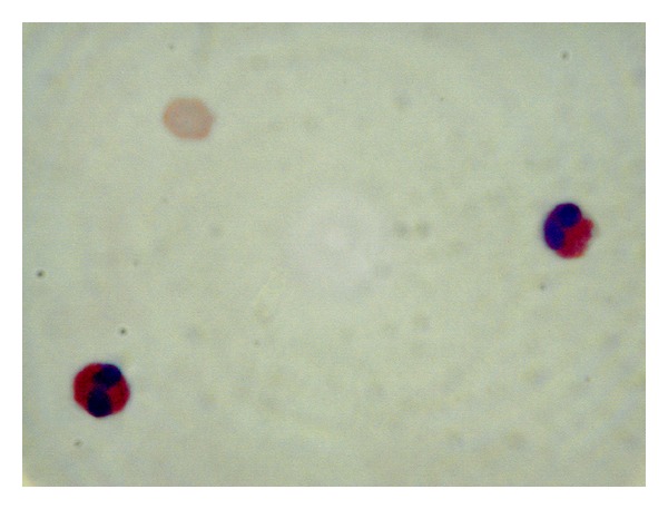
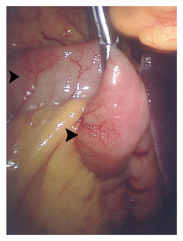
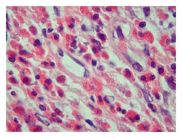
Eosinophilic gastroenteritis is a rare condition characterized by recurrent eosinophilic infiltration of portions of the GI tract and presenting with nonspecific GI symptoms in association with peripheral eosinophilia. Its etiology and pathogenesis remain unclear and its symptoms overlap with many GI and systemic diseases. Thus, both gastroenterologists and general internists need to be aware of this rare condition. We present a case of a 55-year-old male with diffuse abdominal pain and distention for two weeks. His physical examination was significant for moderate ascites. Initial work-up demonstrated severe peripheral blood eosinophilia, normal liver function tests, thickening of the stomach and small bowel wall, and elevated serum IgE. Upper endoscopy and extensive testing for malignancy and parasitic infections failed to establish a diagnosis. Ascitic fluid analysis showed significant eosinophilia. Further, a full-thickness jejunal showed marked eosinophilic infiltration of the serosa and muscularis propria. Subsequent treatment with oral prednisone resulted in normalization of laboratory and radiologic abnormalities in a few week period.
Figures
References
-
- Agostino A, Parenzi A. Eosinophilic gastroenteritis. A case with predominant involvement of mucosal and muscular layers. Minerva Medica. 1994;85(12):655–658. - PubMed
-
- Klein NC, Hargrove RL, Sleisenger MH, Jeffries GH. Eosinophilic gastroenteritis. Medicine. 1970;49(4):299–319. - PubMed
-
- Lee M, Hodges WG, Huggins TL, Lee EL. Eosinophilic gastroenteritis. Southern Medical Journal. 1996;89(2):189–194. - PubMed
-
- Durieu I, Nove-Josserand R, Cathebras P, Vital Durand D, Rousset H, Levrat R. Eosinophilic ascites. Report of two cases. Revue de Medecine Interne. 1992;13(6):446–448. - PubMed
Publication types
LinkOut - more resources
Full Text Sources
