. 2012 Jul 11;134(27):11261-8.
doi: 10.1021/ja3038273. Epub 2012 Jun 29.

Single-molecule Förster resonance energy transfer reveals an innate fidelity checkpoint in DNA polymerase I

Affiliations

Single-molecule Förster resonance energy transfer reveals an innate fidelity checkpoint in DNA polymerase I

Svitlana Y Berezhna et al. J Am Chem Soc. .

Abstract

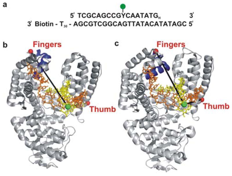
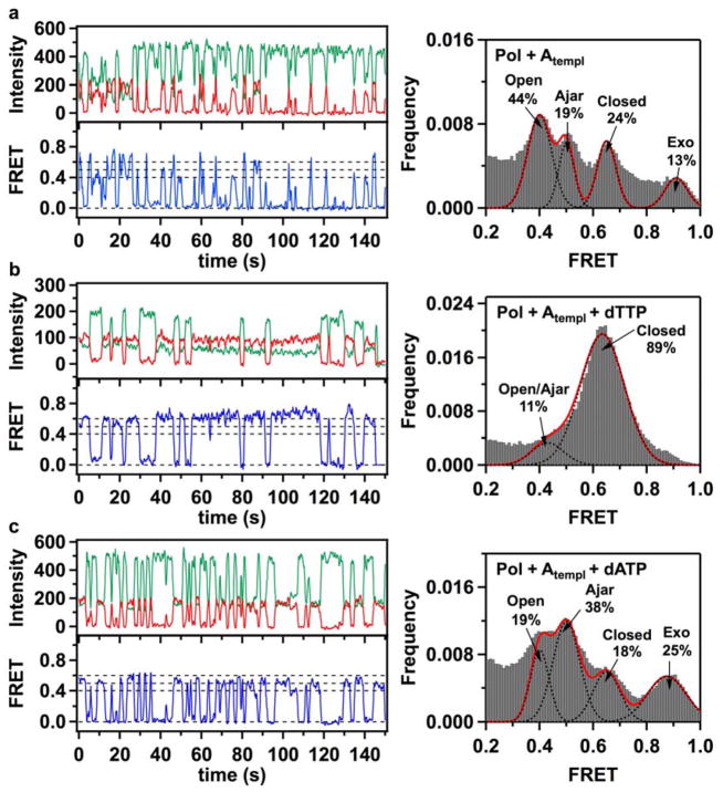
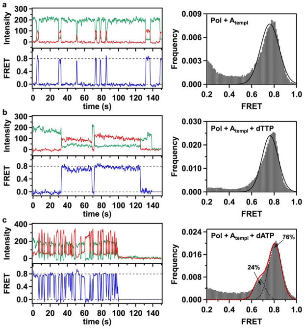
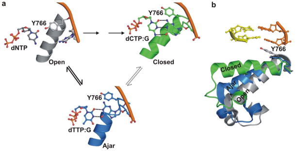
Enzymatic reactions typically involve complex dynamics during substrate binding, conformational rearrangement, chemistry, and product release. The noncovalent steps provide kinetic checkpoints that contribute to the overall specificity of enzymatic reactions. DNA polymerases perform DNA replication with outstanding fidelity by actively rejecting noncognate nucleotide substrates early in the reaction pathway. Substrates are delivered to the active site by a flexible fingers subdomain of the enzyme, as it converts from an open to a closed conformation. The conformational dynamics of the fingers subdomain might also play a role in nucleotide selection, although the precise role is currently unknown. Using single-molecule Förster resonance energy transfer, we observed individual Escherichia coli DNA polymerase I (Klenow fragment) molecules performing substrate selection. We discovered that the fingers subdomain actually samples through three distinct conformations--open, closed, and a previously unrecognized intermediate conformation. We measured the overall dissociation rate of the polymerase-DNA complex and the distribution among the various conformational states in the absence and presence of nucleotide substrates, which were either correct or incorrect. Correct substrates promote rapid progression of the polymerase to the catalytically competent closed conformation, whereas incorrect nucleotides block the enzyme in the intermediate conformation and induce rapid dissociation from DNA. Remarkably, incorrect nucleotide substrates also promote partitioning of DNA to the spatially separated 3'-5' exonuclease domain, providing an additional mechanism to prevent misincorporation at the polymerase active site. These results reveal the existence of an early innate fidelity checkpoint, rejecting incorrect nucleotide substrates before the enzyme encloses the nascent base pair.


Figures

Figure 1
Figure 1. Schematic illustration of labeling strategy used to probe the finger-closing conformational change in Pol I KF, based on crystal structures of homolog Bacillus stearothermophilus (Bst) Pol I
(a) The sequence of the primer-template DNA used in the experiments. The primer strand was dideoxy terminated at the 3′ end (denoted by a subscript H) to prevent nucleotide incorporation. The Y represents an amino-dT used as the labeling site for the Alexa-Fluor 488 donor dye. The template strand is 3′ biotin labeled for surface attachment. The T30 extension at the 3′ end of the template strand separates the duplex portion of the primer/template from the surface. (b) The open polymerase-DNA binary complex (PDB code 1L3S). (c) The closed polymerase-DNA-dNTP ternary complex (PDB code 1LV5). The mobile segment of the protein including the O-helix (residues 680–714 in Bst Pol I, corresponding to residues 732–766 in KF Pol I) is highlighted in dark blue. The yellow and orange oligonucleotides are the primer and template strands, respectively. The red spheres represent positions of the Alexa-Fluor 594 acceptor, attached respectively either to amino acid 744 (L744C KF) in the fingers-labeled construct or to residue 550 (K550C KF) in the thumb-labeled construct (Pol I KF residue numbers). The green sphere represents the labeling position of the donor on the DNA primer.
Figure 2
Figure 2. Fluorescence intensity time traces, smFRET efficiency trajectories and FRET efficiency histograms for finger-labeled polymerase molecules (L744C KF)
(a) Binary Pol+Atempl complexes, formed by binding of acceptor-labeled L744C KF molecules to donor-labeled DNA molecules with A in the template extension position, show fast sampling of three bound states, with mean FRET efficiencies of 0.41, 0.50 and 0.63. A small fraction of complexes populate a separated 0.90 FRET state. The assignment of the individual peaks to open, ajar and closed conformations of the fingers subdomain, and to a population of DNA bound at the 3′-5′ exonuclease (exo) site, are discussed in the text. (b) Correct ternary Pol+Atempl+dTTP complexes, formed in the presence of 1 mM dTTP in the solution, show stable binding in the 0.63 FRET state (left). Two separate peaks in the lower FRET region are not resolved and appear as a shoulder at 0.45 FRET, while a higher 0.9 FRET state is no longer apparent (right). (c) In the presence of 1 mM incorrect dATP nucleotides, Pol I KF frequently samples short-lived 0.4 and 0.5 FRET states, with decreased number of molecules in the 0.63 FRET state. The number of complexes populating the 0.9 FRET state increases. In all cases, the green and red traces show background corrected fluorescence intensities in the donor and acceptor channels, respectively. The blue lines show corresponding smFRET trajectories. The dashed lines positioned at 0, 0.4, 0.5 and 0.6 FRET efficiencies are for ease of visual inspection. Histograms of smFRET efficiencies compiled from multiple trajectories (right) were fitted using multiple Gaussian functions. Dashed black lines show individual Gaussian fits, with the red line corresponding to a composite sum of Gaussians. The percentage numbers on each graph indicate the fraction of complexes in different populations, obtained from the peak areas of the Gaussian fits. The numbers of individual smFRET time trajectories used to construct each histogram were 320 (a), 244 (b) and 299 (c).
Figure 3
Figure 3. Fluorescence time traces, smFRET efficiency trajectories and FRET efficiency histograms for thumb-labeled polymerase molecules (K550C KF)
(a) Binding events of acceptor-labeled K550C KF polymerases to immobilized donor-labeled DNA molecules produced short-lived FRET bursts. A composite FRET efficiency histogram compiled from 228 trajectories shows that binary Pol+Atempl complexes populate a single 0.78 FRET state, in contrast with the three-state behavior of L744C KF. (b) Correct Pol+Atempl+dTTP complexes formed in the presence of 1 mM dTTP in the solution demonstrate that K550C molecules exhibit a prolonged period (exceeding 5 s) in a 0.80 FRET state. The composite histogram is compiled from 208 trajectories. (c) In the presence of 1 mM incorrect dATP nucleotides, K550C KF demonstrates frequent sampling of a 0.81 FRET state, reflecting formation of unstable incorrect ternary Pol+Atempl+dATP complexes. The FRET efficiency histogram (compiled from 228 trajectories) also reveals a sub-population (24%) of complexes captured in a 0.7 FRET state. In all cases, the green, red and blue traces correspond to the donor intensity, acceptor intensity and corresponding FRET efficiency trajectories, respectively. The dashed lines positioned at 0 and 0.8 FRET efficiencies are for ease of visual inspection. Gaussian fits to the histogram peaks are shown as black lines. The red line in panel c represents a composite sum of the two fitted Gaussian peaks.
Figure 4
Figure 4. Proposed reaction pathway steps at Pol I KF active site preceding phosphodiester bond formation
The model is based on the results of the present smFRET study and crystal structures of the close KF homologue Bst Pol I,. (a) In the initial step, the complex of polymerase, DNA and incoming dNTP adopts an open conformation of the fingers (left). The structure shown is based on the binary complex of Bst Pol I (PDB file 1L3U), with a dNTP positioned adjacent to the O-helix by model building. Only the templating base, incoming dNTP, O-helix and conserved tyrosine residue (Tyr766 in Pol I KF) are shown. The open conformation exists in equilibrium with ajar and closed conformations. If the nucleotide substrate is correct, the ternary complex rapidly progresses to the closed conformation. The resulting structure (upper right) is based on the closed complex of Bst Pol I with dCTP paired with template dG (PDB code 1LV5). The intermediate ajar conformation of the fingers is only fleetingly populated in our experiments when the correct dNTP is present. In contrast, the polymerase is largely blocked in the ajar conformation when an incorrect nucleotide is bound (lower center). The structure shown is based on a ternary complex of Bst Pol I with dTTP mispaired with template dG (PDB code 3HP6). In this structure, the template base has rotated from the pre-insertion to insertion site, allowing mispairing with the incoming dNTP. (b) An overlay of three crystal structures of Bst Pol I provides a comparison of the fingers subdomain positions in open, ajar and closed conformations ,. The O helix of the fingers subdomain (encompassing amino residues 732–766 in KF) is shown in ribbons, except for the conserved tyrosine and two post-insertion site DNA base pairs, which are shown as sticks. Incoming nucleotides have been omitted from the ajar and closed conformations in panel (b) for clarity. All models were generated using PyMol.

Similar articles

Cited by

References

    1. Kunkel TA. J Biol Chem. 2004;279:16895. - PubMed
    1. Dahlberg ME, Benkovic SJ. Biochemistry. 1991;30:4835. - PubMed
    1. Eger BT, Benkovic SJ. Biochemistry. 1992;31:9227. - PubMed
    1. Wong I, Patel SS, Johnson KA. Biochemistry. 1991;30:526. - PubMed
    1. Joyce CM, Benkovic SJ. Biochemistry. 2004;43:14317. - PubMed

Publication types

MeSH terms