

The role of saposin C in Gaucher disease
- PMID: 22652185
- PMCID: PMC3534739
- DOI: 10.1016/j.ymgme.2012.04.024
The role of saposin C in Gaucher disease
Abstract
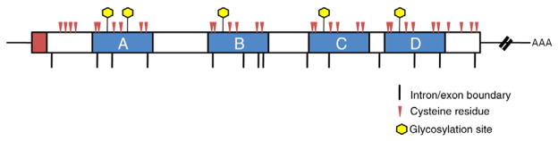
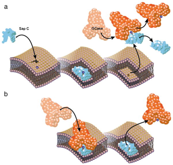
Saposin C is one of four homologous proteins derived from sequential cleavage of the saposin precursor protein, prosaposin. It is an essential activator for glucocerebrosidase, the enzyme deficient in Gaucher disease. Gaucher disease is a rare autosomal recessive lysosomal storage disorder caused by mutations in the GBA gene that exhibits vast phenotypic heterogeneity, despite its designation as a "simple" Mendelian disorder. The observed phenotypic variability has led to a search for disease modifiers that can alter the Gaucher phenotype. The PSAP gene encoding saposin C is a prime candidate modifier for Gaucher disease. In humans, saposin C deficiency due to mutations in PSAP results in a Gaucher-like phenotype, despite normal in vitro glucocerebrosidase activity. Saposin C deficiency has also been shown to modify phenotype in one mouse model of Gaucher disease. The role of saposin C as an activator required for normal glucocerebrosidase function, and the consequences of saposin C deficiency are described, and are being explored as potential modifying factors in patients with Gaucher disease.
Published by Elsevier Inc.
Conflict of interest statement
The authors declare no conflicts of interest.
Figures

Similar articles
-
Phenotype Expansion for Atypical Gaucher Disease Due to Homozygous Missense PSAP Variant in a Large Consanguineous Pakistani Family.Genes (Basel). 2022 Apr 9;13(4):662. doi: 10.3390/genes13040662. Genes (Basel). 2022. PMID: 35456468 Free PMC article.
-
Non-neuronopathic Gaucher disease due to saposin C deficiency.Clin Genet. 2007 Dec;72(6):538-42. doi: 10.1111/j.1399-0004.2007.00899.x. Epub 2007 Oct 7. Clin Genet. 2007. PMID: 17919309
-
A Type 3 Gaucher-Like Disease Due To Saposin C Deficiency in Two Emirati Families Caused by a Novel Splice Site Variant in the PSAP Gene.J Mol Neurosci. 2022 Jun;72(6):1322-1333. doi: 10.1007/s12031-022-01987-y. Epub 2022 Mar 22. J Mol Neurosci. 2022. PMID: 35316504
-
Mutations causing Gaucher disease.Hum Mutat. 1994;3(1):1-11. doi: 10.1002/humu.1380030102. Hum Mutat. 1994. PMID: 8118460 Review.
-
Involvement of Gaucher Disease Mutations in Parkinson Disease.Curr Protein Pept Sci. 2017;18(7):758-764. doi: 10.2174/1389203717666160311115956. Curr Protein Pept Sci. 2017. PMID: 26965692 Review.
Cited by
-
Engineering of Saposin C Protein Chimeras for Enhanced Cytotoxicity and Optimized Liposome Binding Capability.Pharmaceutics. 2021 Apr 19;13(4):583. doi: 10.3390/pharmaceutics13040583. Pharmaceutics. 2021. PMID: 33921905 Free PMC article.
-
Prosaposin variants in sporadic, familial, and early-onset Parkinson's disease: a Taiwanese case-control study and meta-analysis.Sci Rep. 2024 Jan 26;14(1):2225. doi: 10.1038/s41598-024-51646-y. Sci Rep. 2024. PMID: 38278831 Free PMC article.
-
Glucocerebrosidase is shaking up the synucleinopathies.Brain. 2014 May;137(Pt 5):1304-22. doi: 10.1093/brain/awu002. Epub 2014 Feb 14. Brain. 2014. PMID: 24531622 Free PMC article. Review.
-
Intracellular Proteolysis of Progranulin Generates Stable, Lysosomal Granulins that Are Haploinsufficient in Patients with Frontotemporal Dementia Caused by GRN Mutations.eNeuro. 2017 Aug 18;4(4):ENEURO.0100-17.2017. doi: 10.1523/ENEURO.0100-17.2017. eCollection 2017 Jul-Aug. eNeuro. 2017. PMID: 28828399 Free PMC article.
-
Protective Effect of the LRRK2 Kinase Inhibition in Human Fibroblasts Bearing the Genetic Variant GBA1 K198E: Implications for Parkinson's Disease.Neuromolecular Med. 2025 May 21;27(1):42. doi: 10.1007/s12017-025-08864-y. Neuromolecular Med. 2025. PMID: 40397198 Free PMC article.
References
-
- Kishimoto Y, Hiraiwa M, O’Brien JS. Saposins: structure, function, distribution, and molecular genetics. J Lipid Res. 1992;33:1255–1267. - PubMed
-
- Vaccaro AM, Salvioli R, Tatti M, Ciaffoni F. Saposins and their interaction with lipids. Neurochem Res. 1999;24:307–314. - PubMed
-
- Futerman AH, Zimran A. Gaucher disease. CRC/Taylor & Francis; Boca Raton: 2006.
-
- Hiraiwa M, Martin BM, Kishimoto Y, Conner GE, Tsuji S, O’Brien JS. Lysosomal proteolysis of prosaposin, the precursor of saposins (sphingolipid activator proteins): its mechanism and inhibition by ganglioside. Arch Biochem Biophys. 1997;341:17–24. - PubMed
Publication types
MeSH terms
Substances
Grants and funding
LinkOut - more resources
Full Text Sources
Other Literature Sources
Medical
Miscellaneous
