Review
. 2012 Jun 1;17(6):1996-2023.
doi: 10.2741/4034.

Protein-mediated enamel mineralization

Affiliations
Review

Protein-mediated enamel mineralization

Janet Moradian-Oldak. Front Biosci (Landmark Ed). .

Abstract

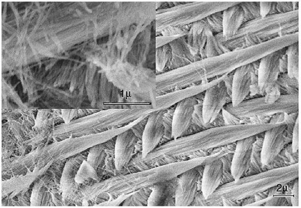
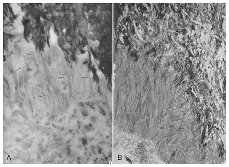
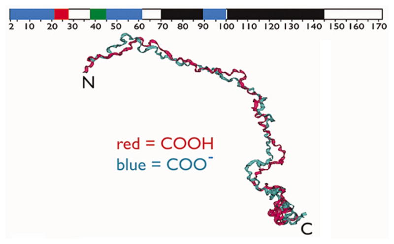
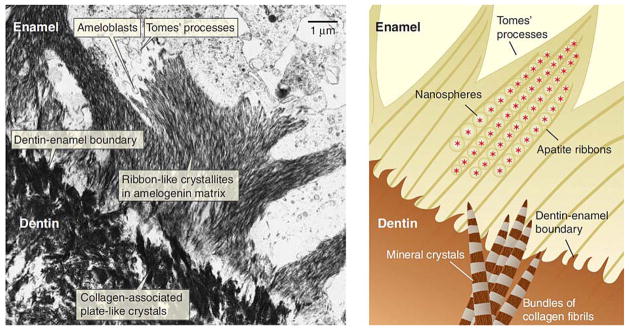
Enamel is a hard nanocomposite bioceramic with significant resilience that protects the mammalian tooth from external physical and chemical damages. The remarkable mechanical properties of enamel are associated with its hierarchical structural organization and its thorough connection with underlying dentin. This dynamic mineralizing system offers scientists a wealth of information that allows the study of basic principels of organic matrix-mediated biomineralization and can potentially be utilized in the fields of material science and engineering for development and design of biomimetic materials. This chapter will provide a brief overview of enamel hierarchical structure and properties and the process and stages of amelogenesis. Particular emphasis is given to current knowledge of extracellular matrix protein and proteinases, and the structural chemistry of the matrix components and their putative functions. The chapter will conclude by discussing the potential of enamel for regrowth.


Figures

Figure 1
Figure 1
Scanning electron micrograph of etched surface of mouse incisor tooth enamel showing the interwoven arrangement of the prisms and inter-prismatic material. Inset: Higher magnification SEM micrograph showing the calcium fluoridated carbonated apatite crystals arranged in parallel boundless within the prisms.
Figure 2
Figure 2
(A) Ameloblast membrane projections on dentin surface in human teeth. Detailed picture of the distal part of the ameloblast cell. Filaments with cross banding typical of collagen are located between the narrow processes extending from the distal surface of the ameloblast cell toward the dentine layer. Magnification=× 67,000. Reproduced with permission from reference # ; Elsevier through Copyright Clearance Center. (B) The newly formed enamel shows a very irregular wavy boundary at the enamel-dentin junction, EDJ. Processes from enamel and dentine interdigitate, magnification=X 39,000. Reproduced with permission from reference # ; Elsevier through Copyright Clearance Center. D: dentin, C: collagen, EDJ: Enamel Dentin Junction, A: Ameloblasts, PM: Plasma membrane of the ameloblasts.
Figure 3
Figure 3
Backbone aligned best structures obtained for the fully protonated (Red) and deprotonated (Blue) forms of recombinant porcine amelogenin (rP172) in monomeric form, showing that the molecule is extended (backbone rmsd = 4.7 Å). The ribbon on the top represents secondary structure assignment based upon nOe and 3J data in this figure; black = PPII, red = alpha-helix, blue = extended beta strand, green = beta turn or loop, and white = random coil or unstructured. Reproduced with permission from (58).
Figure 4
Figure 4
(A) Micrograph of the dentin-enamel boundary in a developing tooth. The apatite of dentin (lower left) forms nanometer-scale plates that initially grow individually and independently in the gap spaces along the collagen fibrils. The ribbon-like apatite crystallites of the enamel (top right) form within the amelogenin-rich enamel matrix. (B) A close-up view (not to scale) of the hypothesized mineral deposition processes in dentin and enamel near the dentin-enamel boundary. In the dentin, plate-like apatite crystals grow in the periodic gap spaces along the collagen fibrils and fibril bundles. The apatite crystal c axis is mostly aligned with the long axis of the collagen fibril. In the enamel, Du et al. (72) propose that linear aggregates of polarized, self-assembled amelogenin nanospheres form a negatively charged template that induces apatite formation. Reproduced with permission from reference # ; The American Association for the Advancement of Science through Copyright Clearance Center.
Figure 5
Figure 5
Suggested mechanism for in vitro hierarchically organized, elongated HAP microstructures formed by co-assembly of amelogenin and calcium phosphate nanoclusters, based on in vitro experiments Reproduced with permission from (109).
Figure 6
Figure 6
SEM images of fractured incisors showing the defects in enamel formation of amelogenin (Amel), ameloblastin (Ambn), and enamelin (Enam) null-mice. (A) wild type mouse. (B) the enamel from the null mouse does not have a normal prismatic structure and is markedly reduced in thickness compared with that of the wild type mouse shown at the same magnification as A, Bars in A and B = 10 micrometer. Reproduced with permission from reference #. (C) Heterezygote Ambn+/− and (D) Homozygote Ambn −/− mice revealing a displastic layer with a rough surface over dentin and lack of a prism pattern in D. Reproduced with permission from reference #. (E) Heterezygote Enam −/+ and (F) Homozygote Enam −/ − mice. The forming enamel in the Enam−/ − mouse (F), is unmineralized, but shows remarkable similarity to normal enamel matrix structure. Reproduced with permission from (131). E: Enamel, D: Dentin, dE: defective enamel, Am: Ameloblasts, Od: Odontoblasts; arrowheads in E and F indicate the dentinoenamel junction.
Figure 7
Figure 7
SEM images of octacalcium phosphate crystals grown in the cation selective membrane system in the presence of (A) 40 microgram/mL 32-kDa enamelin, (B) 40 microgram/mL 32-kDa enamelin in 10% rP148 porcine amelogenin. Note the increase in crystals aspect ratios in B when compared to A. (C) Computer simulation models for OCP crystals grown in the presence of different amounts of enamelin (Length, width, and thickness values are based on the average values in reference #137).
Figure 8
Figure 8
(A) Categorization plot of mean pH (solid squares) ± SD (lines) and SEM (open boxes; n = 10 teeth) for enamel strips cut at 0.5-mm length across the secretory (S) and maturation (M) stages of amelogenesis (R = location of the molar reference as described by Smith et al (161)). The average measured pH of enamel strips remains fairly constant across the secretory stage (S) and into the early maturation stage (M). Reproduced with permission from reference #. (B and C) Cyclical changes in pH in developing bovine incisor: (B) Universal indicator color standards at different pH values used to stain bovine incisor shown in C. (C) A bovine developing tooth cut in half sagittally. The left half was stained with a pH (Universal) indicator mixture for l-2 min. Alternate stripes of orange correspond to pH 5.5–6.0 and of green to pH 7.0. The right half was stained with GBHA (a calcium chelator dye) solution and rinsed with ethanol. Red stripes of staining correspond to the neutral bands of green staining with the pH indicator and unstained white zones to acidic orange zones. Orange or green coloration occured not only on the forming surface but also in depth on enamel. Reproduced with permission from (98)
Figure 9
Figure 9
SEM images of fluoridated apatite–amelogenin coatings prepared on enamel surface (longitudinal sections) with 0 (A) 20 (micrograms/mL (B) 33 micrograms/mL (C) of rP172 amelogenin in the mineralization solution and 1 mg/L F. All the images were taken under the same magnification (204).

References

    1. Lowenstam HA, Weiner S. On Biomineralization. Oxford University Press; New York: 1989.
    1. Moradian-Oldak J, Paine ML. Mammalian Enamel Formation. In: Sigel Astrid, Sigel H, Sigel RKO., editors. Metal Ions In Life Sciences. John Wiley & Sons, Ltd; Chichester: 2008.
    1. Fincham A, Moradian-Oldak J, Simmer J. The structural biology of the developing dental enamel matrix. J Struct Biol. 1999;126(3):270–99. - PubMed
    1. Smith C. Cellular and chemical events during enamel maturation. Crit Rev Oral Biol Med. 1998;9:128–161. - PubMed
    1. Robinson C, Kirkham J, Brookes SJ, Bonass WA, Shore RC. The chemistry of enamel development. Int J Dev Biol. 1995;39(1):145–52. - PubMed

Publication types

MeSH terms

LinkOut - more resources