Review
. 2012 Jun 1;17(6):2197-212.
doi: 10.2741/4045.

The Fbw7 and betaTRCP E3 ubiquitin ligases and their roles in tumorigenesis

Affiliations
Review

The Fbw7 and betaTRCP E3 ubiquitin ligases and their roles in tumorigenesis

Alan W Lau et al. Front Biosci (Landmark Ed). .

Abstract

The Ubiquitin Proteasome System (UPS) is a major regulator of protein abundance in the cell. The UPS influences the functions of multiple biological processes by targeting key regulators for destruction. E3 ubiquitin ligases are a vital component of the UPS machinery, working with E1 and E2 enzymes to bind substrates and facilitate the transfer of ubiquitin molecules onto the target protein. This poly-ubiquitination, in turn, directs the modified proteins for proteolysis by the 26S proteasome. As the UPS regulates the degradation of multiple oncogenes and tumor suppressors, the dysregulation of this pathway is known to promote various diseases including cancer. While E1 and E2 enzymes have only been minimally linked to cancer development, burgeoning amounts of evidence have implicated loss or gain of E3 function as a key factor in cancer initiation and progression. This review will examine the literature on two SCF-type E3 ligases, SCFFbw7 and SCFbeta-TRCP. In particular, we will highlight novel substrates recently identified for these two E3 ligases, and further discuss how UPS regulation of these targets may promote carcinogenesis.


Figures

Figure 1
Figure 1
Illustration of the Ubiquitination Reaction Pathways. Initially, the E1 enzyme activates, in an ATP-dependent manner, the 76 amino acid ubiquitin molecule by forming a high-energy thiol ester bond with ubiquitin. This activated molecule is then transferred to the E2 conjugating enzyme. The E3 ligase then positions the target substrate near the E2 enzyme allowing for the transfer of ubiquitin. In the case of HECT E3 ligases however, the E3 ligase directly transfers the ubiquitin molecule onto the target substrate. Once a chain of four or more ubiquitin molecules is placed onto the substrate protein, the molecule is then targeted for proteolysis by the 26 proteasome.
Figure 2
Figure 2
Structural Illustration of the SCF Family of E3 ligases. An SCF-type E3-ligase is a multi-subunit complex consisting of three invariable subunits and one variable subunit. The three static subunits include a catalytic RING subunit (Rbx1) that interacts with the E2, a scaffolding subunit (Cul1), and an adaptor subunit (Skp1). The variable molecule is known as the F-box protein (FBP). The major function of the F-box protein is to recruit specific substrates to the E3 complex via substrate interaction domains. The two largest classes of interaction domains found on FBPs are WD40 repeats and leucine-rich repeats (LRRs). A third type of FBP also exists which contains neither WD40 repeats nor LRRs. This third class of F-box proteins contains other types of interaction domains or no recognizable domain at all. To date, there have been approximately 69 FBPs identified in the human genome. Furthermore, unlike HECT E3 ligases that can directly conjugate ubiquitin onto the target, SCF complexes bridge the interaction between the E2 enzyme and the substrate.
Figure 3
Figure 3
Illustration of the various types of E3 ligases and F-box proteins. E3 ubiquitin ligases are categorized into three major classes: U-box-type, Ring-finger-type, and HECT-type. U-box- and Ring-finger-type ligases function by bridging the interaction between the E2 enzyme and the substrate. HECT-type ligases are capable of directly conjugating ubiquitin onto the target protein. Ring-finger E3s are further divided into subfamilies including those that are cullin-based as well as the anaphase-promoting complex/cyclosome (APC/C). The SCF complex is a Cul1-based E3 ligase that consists of four components, Skp1, Cul1, Rbx1, and a variable subunit F-box protein. The F-box protein serves as a substrate recognition subunit for the SCF ligase by interacting with target proteins via its c-terminal binding domains. These F-box proteins fall into three major classes based on the substrate interaction domain that is present. Categories include F-box proteins that contain WD40 repeats (FBXW), leucine rich repeats (FBXL), or other protein interaction domains (FBXO).
Figure 4
Figure 4
Fbw7 regulates cellular apoptosis by targeting the pro-survival factor Mcl-1 for degradation. (A) Loss of Fbw7 function leads to elevated levels of its many substrates including c-Myc, c-Jun, and Notch-1. While over-expression of these oncoproteins do give the cell a growth advantage, they also promote apoptosis, which in theory would inhibit tumorigenesis. Therefore, it was unclear how cells deficient in Fbw7 function can thrive in a setting of upregulated c-Jun, c-Myc or Notch-1. (B) We recently identified the pro-survival factor Mcl-1 as a novel target of SCFFbw7. Upon loss of Fbw7 function, cellular Mcl-1 levels are elevated. Higher expression of Mcl-1 in turn suppresses apoptosis caused by enhanced expression of c-Myc, c-Jun or Notch-1. This could then allow for uncontrolled cell growth and tumorigenesis.
Figure 5
Figure 5
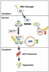
Multi-site phosphorylation of Mdm2 by CKI in response to DNA damage promotes Mdm2 ubiquitination and destruction by SCFbeta-TRCP. In unstressed cells, p53 levels are maintained due to its interaction with Mdm2, which promotes the ubiquitination and proteolysis of p53. Upon DNA damage, CKI translocates to the nucleus where it multi-site phosphorylates Mdm2. This modified Mdm2 species is then recognized and bound by SCFbeta-TRCP. The E3-ligase then in turn poly-ubiquitinates Mdm2 and promotes its degradation by the 26 proteasome.

References

    1. Komander D. The emerging complexity of protein ubiquitination. Biochem Soc Trans. 2009;37:937–53. - PubMed
    1. Hochstrasser M. Ubiquitin-dependent protein degradation. Annu Rev Genet. 1996;30:405–39. - PubMed
    1. Nakayama KI, Nakayama K. Regulation of the cell cycle by SCF-type ubiquitin ligases. Semin Cell Dev Biol. 2005;16:323–33. - PubMed
    1. Mukhopadhyay D, Riezman H. Proteasome-independent functions of ubiquitin in endocytosis and signaling. Science. 2007;315:201–5. - PubMed
    1. Thrower JS, Hoffman L, Rechsteiner M, Pickart CM. Recognition of the polyubiquitin proteolytic signal. Embo J. 2000;19:94–102. - PMC - PubMed

Publication types

MeSH terms

Substances