Review
. 2012 Jul 7;4(13):3833-42.
doi: 10.1039/c2nr31040f. Epub 2012 Jun 1.

Graphene: a versatile nanoplatform for biomedical applications

Affiliations
Review

Graphene: a versatile nanoplatform for biomedical applications

Yin Zhang et al. Nanoscale. .

Abstract

Graphene, with its excellent physical, chemical, and mechanical properties, holds tremendous potential for a wide variety of biomedical applications. As research on graphene-based nanomaterials is still at a nascent stage due to the short time span since its initial report in 2004, a focused review on this topic is timely and necessary. In this feature review, we first summarize the results from toxicity studies of graphene and its derivatives. Although literature reports have mixed findings, we emphasize that the key question is not how toxic graphene itself is, but how to modify and functionalize it and its derivatives so that they do not exhibit acute/chronic toxicity, can be cleared from the body over time, and thereby can be best used for biomedical applications. We then discuss in detail the exploration of graphene-based nanomaterials for tissue engineering, molecular imaging, and drug/gene delivery applications. The future of graphene-based nanomaterials in biomedicine looks brighter than ever, and it is expected that they will find a wide range of biomedical applications with future research effort and interdisciplinary collaboration.


Figures

Fig. 1
Fig. 1
Tissue engineering with graphene. (a) A schematic diagram depicting growth and differentiation of human neural stem cells (hNSCs) on graphene coated with laminin. (b) A bright-field image of hNSCs at the boundary area between glass (left) and graphene (right) at 10 h after cell seeding. (c) Bright-field (left) and immunofluorescence (right) images of hNSCs at 5 days after seeding. Green: nestin (a marker for hNSCs); Blue: DAPI (nuclei). All scale bars represent 200 μm. Adapted from ref. .
Fig. 2
Fig. 2
Optical imaging of graphene-based nanomaterials. (a) Cellular uptake of folic acid-conjugated QD-rGO in human breast cancer MCF-7 and HeLa cells, where QD fluorescence is shown in red-orange. (b) A schematic representation of Cy7-labeled GO through six-arm branched PEG chains. (c) In vivo fluorescence imaging of mice bearing different tumors (indicated by arrows) after intravenous injection of Cy7-labeled GO. Adapted from ref. and .
Fig. 3
Fig. 3
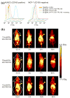
Positron emission tomography (PET) imaging of radiolabeled GO. (a) Flow cytometry analysis of GO conjugates in CD105 positive human umbilical vein endothelial cells (HUVECs) and CD105 negative MCF-7 cells. (b) Serial PET imaging of 4T1 tumor-bearing mice after intravenous injection of NOTA-GO-TRC105, labeled with each of the three isotopes: 61Cu, 66Ga, and 64Cu. TRC105 is an antibody that binds to CD105. Arrowheads indicate the tumors. Adapted from ref. .
Fig. 4
Fig. 4
In vivo multimodality imaging of rGO-IONP in 4T1 tumor-bearing mice. (a) Serial fluorescence imaging of Cy5-labeled rGO-IONP after intravenous injection. Yellow arrowheads indicated the tumor. (b) T2-weighted magnetic resonance imaging of rGO-IONP, where the red circles indicated the tumors. (c) Photoacoustic (PA) imaging of rGO-IONP. Adapted from ref. .
Fig. 5
Fig. 5
Synergistic effect of chemo-photothermal therapy with PEGylated graphene oxide. (a) Tumor growth curves of mice in different treatment groups. (b) Mean body weight of mice in different groups after treatment. (c) Representative photos of mice after different treatment. NGO: nanographene oxide; NIR: near-infrared photothermal therapy; DOX: doxorubicin. Adapted from ref. .

References

    1. Lanone S, Boczkowski J. Curr Mol Med. 2006;6:651–663. - PubMed
    1. Gonsalves K, Halberstadt C, Laurencin CT, Nair L. Biomedical Nanostructures. John Wiley & Sons, Inc; Hoboken, New Jersey: 2007.
    1. Wang X, Liu LH, Ramstrom O, Yan M. Exp Biol Med (Maywood) 2009;234:1128–1139. - PMC - PubMed
    1. Cai W, Hsu AR, Li ZB, Chen X. Nanoscale Res Lett. 2007;2:265–281. - PMC - PubMed
    1. Cai W, Hong H. Am J Nucl Med Mol Imaging. 2012;2:136–140. - PMC - PubMed

Publication types