. 2012 Nov;19(11):1836-46.
doi: 10.1038/cdd.2012.67. Epub 2012 Jun 1.

C/EBP homologous protein contributes to cytokine-induced pro-inflammatory responses and apoptosis in β-cells

Affiliations

C/EBP homologous protein contributes to cytokine-induced pro-inflammatory responses and apoptosis in β-cells

F Allagnat et al. Cell Death Differ. 2012 Nov.

Abstract

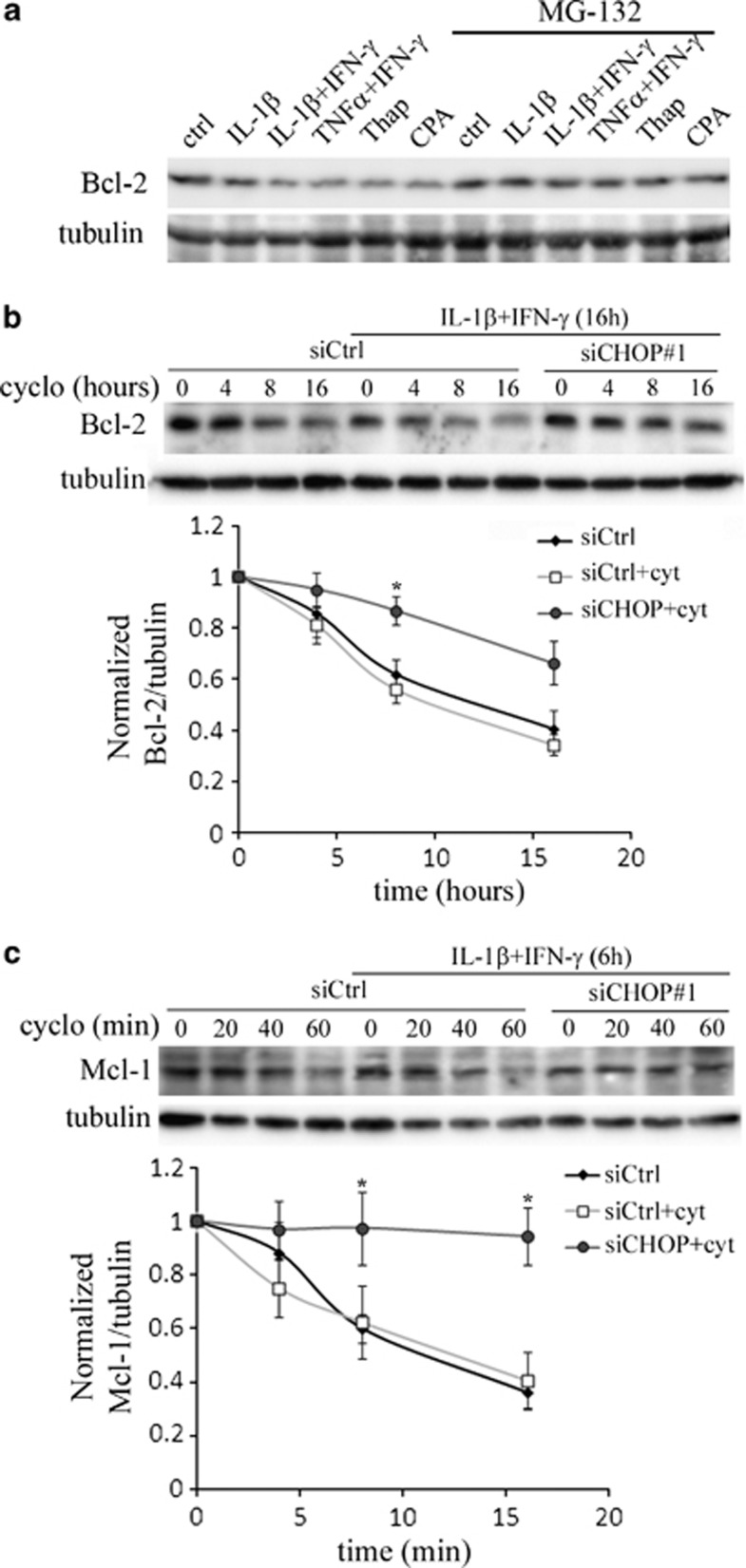
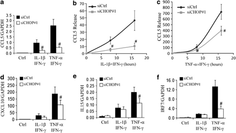
Induction of the C/EBP homologous protein (CHOP) is considered a key event for endoplasmic reticulum (ER) stress-mediated apoptosis. Type 1 diabetes (T1D) is characterized by an autoimmune destruction of the pancreatic β-cells. Pro-inflammatory cytokines are early mediators of β-cell death in T1D. Cytokines induce ER stress and CHOP overexpression in β-cells, but the role for CHOP overexpression in cytokine-induced β-cell apoptosis remains controversial. We presently observed that CHOP knockdown (KD) prevents cytokine-mediated degradation of the anti-apoptotic proteins B-cell lymphoma 2 (Bcl-2) and myeloid cell leukemia sequence 1 (Mcl-1), thereby decreasing the cleavage of executioner caspases 9 and 3, and apoptosis. Nuclear factor-κB (NF-κB) is a crucial transcription factor regulating β-cell apoptosis and inflammation. CHOP KD resulted in reduced cytokine-induced NF-κB activity and expression of key NF-κB target genes involved in apoptosis and inflammation, including iNOS, FAS, IRF-7, IL-15, CCL5 and CXCL10. This was due to decreased IκB degradation and p65 translocation to the nucleus. The present data suggest that CHOP has a dual role in promoting β-cell death: (1) CHOP directly contributes to cytokine-induced β-cell apoptosis by promoting cytokine-induced mitochondrial pathways of apoptosis; and (2) by supporting the NF-κB activation and subsequent cytokine/chemokine expression, CHOP may contribute to apoptosis and the chemo attraction of mononuclear cells to the islets during insulitis.


Figures

Figure 1
Figure 1
CHOP knockdown protects against cytokine-induced β-cell apoptosis. (a) Representative time-course experiment of CHOP protein expression in INS-1E cells treated with IL-1β+IFN-γ (left panel) or TNF-α+IFN-γ (right panel). (b) Time-course analyses of CHOP mRNA expression in human islets exposed to TNF-α+IFN-γ (squares) or IL-1β+IFN-γ (circles). Data are means±S.E.M. of three to five independent experiments. *P<0.05, **P<0.01 versus non-treated condition. (c) Representative western blot of CHOP over tubulin expression in INS-1E untransfected (NT) or transfected with a control siRNA (siCtrl), or two different CHOP siRNA (siCHOP#1 and #2) and treated (+) or not (−) for 15 h with CPA. Data are means±S.E.M. of four independent experiments. (d) INS-1E cells were untransfected (NT, black bars), transfected with siCtrl (stripe bars), siCHOP#1 (white bars), or siCHOP#2 (dotted bars), and treated for 15 h with IL-1β+IFN-γ, TNF-α+IFN-γ or CPA as indicated. (e and f) FACS-purified primary β-cells were transfected with the siCtrl (black bars) or siCHOP#1 (white bars), and treated for 24 h with IL-1β+IFN-γ or TNF-α+IFN-γ. (e) Real-time PCR analyses of CHOP over GAPDH mRNA expression. (d and f) Prevalence of apoptosis was evaluated by HO-PI staining. (d and f) Data are mean±S.E.M. of at least four independent experiments. *P<0.05, **P<0.01 versus respective non-treated condition. #P<0.05, ##P<0.01 versus respective untransfected and siCtrl-transfected condition. (g) Dispersed human islet cells were transfected with the siCtrl, the human siCHOP#1, or the human siCHOP#2 and treated for 48 h with TNF-α+IFN-γ. Prevalence of apoptosis was evaluated by HO-PI staining and expressed as the apoptotic index. Data are mean±S.E.M. of three independent experiments. *P<0.05 versus respective siCtrl-transfected condition
Figure 2
Figure 2
CHOP knockdown protects against cytokine-induced caspase 3 and 9 cleavage. INS-1E cells were transfected with a control siRNA (siCtrl) or siCHOP#1, and treated for 15 h with IL-1β+IFN-γ or TNF-α+IFN-γ. (a) Representative western blot showing CHOP and the cleaved caspase 3 and 9 fragments normalized to the tubulin levels. (b) Quantitative assessment of four independent blots. *P<0.05, **P<0.01 versus respective non-treated condition. #P<0.05 versus respective siCtrl-transfected condition
Figure 3
Figure 3
CHOP knockdown transiently protects against cytokine-induced Mcl-1 and Bcl-2 protein downregulation. (a and b) Western blot analyses of Bcl-2 (a) and Mcl-1 (b) over tubulin in INS-1E cells transfected with a control siRNA (siCtrl) or siCHOP#1, and treated for 15 or 24 h with IL-1β+IFN-γ. Upper panels: representative western blot. Lower panels: quantitative assessment of four independent blots. *P<0.05, **P<0.01 versus respective non-treated condition. #P<0.05 versus respective siCtrl-transfected condition
Figure 4
Figure 4
CHOP knockdown stabilizes Bcl-2 and Mcl-1 proteins in INS-1E cells treated with cytokines. (a) Representative western blot of Bcl-2 expression in INS-1E cells exposed for 15 h to cytokines (IL-1β or TNF-α,±IFN-γ, as indicated), CPA or thapsigargin (thap), with or without the proteasome inhibitor MG-132. Figure is representative of four independent experiments. (b) INS-1E cells were transfected with siCtrl or siCHOP#1, and treated or not for 16 h with IL-1β+IFN-γ. Cells were then further treated or not (time 0) for 4, 8 or 16 h with cycloheximide (30 μM) to inhibit protein synthesis. Western blot analyses of Bcl-2 over tubulin were performed. Upper panel: representative blot. Lower panel: quantitative assessment of four independent blots. The results were normalized by setting the time zero of each condition as 1. *P<0.05 versus respective siCtrl-tranfected cells treated with cytokines. (c) INS-1E cells were transfected with siCtrl or siCHOP#1, and treated or not for 6 h with IL-1β+IFN-γ. The cells were then further treated or not (0) for 20, 40 or 60 min with cycloheximide (30 μM). Western blot analyses of Mcl-1 and tubulin were performed. Upper panel: representative blot. Lower panel: quantitative assessment of four independent blots. The results were normalized by setting the time zero of each condition as 1. *P<0.05 versus respective siCtrl-transfected cells treated with cytokines
Figure 5
Figure 5
CHOP knockdown decreases cytokine-induced NF-κB activity and expression of the NF-κB target genes iNOS and FAS. (a) INS-1E cells were transfected with a NF-κB responsive reporter construct and the control reporter plasmid pRLSV40renilla, together with a control siRNA (black bars) or the CHOP siRNA (white bars), and treated for 8 h with IL-1β+IFN-γ or TNF-α+IFN-γ. Results are mean NF-κB activities±S.E.M. of six independent experiments. (b and c) INS-1E cells were transfected with a NF-κB responsive reporter construct and the control reporter plasmid pRLSV40renilla, together with an empty plasmid (vector) or a plasmid encoding the rat CHOP (pCHOP). (b) Representative western blot of CHOP over tubulin in cells treated or not for 15 h with IL-1β+IFN-γ or TNF-α+IFN-γ, as indicated. (c) NF-κB activity after 8 h of treatment with IL-1β+IFN-γ or TNF-α+IFN-γ. (d) FACS-purified primary β-cells were transfected with the siCtrl (black bars) or siCHOP#1 (white bars), and treated for 24 h with IL-1β+IFN-γ or TNF-α+IFN-γ. Real-time PCR analyses of NOS2 (d) and FAS (e) over GAPDH mRNA expression. Results are means±S.E.M. of four independent experiments. #P<0.05, ##P<0.01 versus respective siCtrl-transfected condition
Figure 6
Figure 6
CHOP knockdown mitigates cytokine-induced expression of inflammation markers and release of CCL5. (a and df) FACS-purified primary β-cells were transfected with the siCtrl (black bars) or siCHOP#1 (white bars), and treated for 24 h with IL-1β+IFN-γ or TNF-α+IFN-γ. Real-time PCR analyses of CCL5 (a), CXCL10 (d), IL15 (e) and IRF7 (f) over GAPDH mRNA expression. (b and c) INS-1E cells were transfected with siCtrl (black lines) or siCHOP#1 (gray lines), and treated for 8 or 15 h with IL-1β+IFN-γ (b) or TNF-α+IFN-γ (c). CCL5 release was evaluated by ELISA of the cell supernatant. All data are means±S.E.M. of at least four independent experiments. #P<0.05 versus respective siCtrl-transfected condition
Figure 7
Figure 7
CHOP knockdown inhibits cytokine-induced p65 translocation and IκB-α degradation. (a and c) INS-1E cells were transfected with siCtrl or siCHOP#1 and treated for 8 h with IL-1β+IFN-γ or TNF-α+IFN-γ. (a) Representative western blot of p65 and CHOP in total extracts (input) or after co-immunoprecipitation using anti-p65, anti-CHOP or anti-HA antibodies. (b) p65 localization was analyzed by immunofluorescence in INS-1E cells. Data are representative of four independent experiments. (c) Upper panel: representative western blot showing CHOP, IKK-β, IκB-β and IκB-α, normalized to the p65 levels. Lower panel: quantitative assessment of IκB-α over p65 expression of four independent blots. *P<0.05 versus respective non-treated condition. #P<0.05 versus respective siCtrl-transfected condition
Figure 8
Figure 8
Scheme depicting the role of CHOP cytokine-induced NF-κB activity and apoptosis. Pro-inflammatory cytokines stimulate NF-κB activity and JNK phosphorylation, leading to NO production and ER stress, and resulting in eIF2α phosphorylation and CHOP expression. In turn, CHOP facilitates the degradation of IκBα, thereby amplifying NF-κB activity. CHOP also contributes to the degradation of the anti-apoptotic proteins Bcl-2 and Mcl-1, leading to activation of the mitochondrial pathway of apoptosis. Furthermore, the impact of CHOP on NF-κB activity may contribute to inflammatory processes triggered by cytokines in β-cells, putatively aggravating insulitis

References

    1. Patterson CC, Dahlquist GG, Gyurus E, Green A, Soltesz G. Incidence trends for childhood type 1 diabetes in Europe during 1989–2003 and predicted new cases 2005–20: a multicentre prospective registration study. Lancet. 2009;373:2027–2033. - PubMed
    1. Todd JA. Etiology of type 1 diabetes. Immunity. 2010;32:457–467. - PubMed
    1. Eizirik DL, Colli ML, Ortis F. The role of inflammation in insulitis and beta-cell loss in type 1 diabetes. Nat Rev Endocrinol. 2009;5:219–226. - PubMed
    1. Eizirik DL, Cardozo AK, Cnop M. The role for endoplasmic reticulum stress in diabetes mellitus. Endocr Rev. 2008;29:42–61. - PubMed
    1. Gurzov EN, Eizirik DL. Bcl-2 proteins in diabetes: mitochondrial pathways of beta-cell death and dysfunction. Trends Cell Biol. 2011;21:424–431. - PubMed

Publication types

MeSH terms