. 2012 Jul;60(7):425-30.
doi: 10.1007/s11748-012-0066-7. Epub 2012 Jun 1.

One-dimensional mean computed tomography value evaluation of ground-glass opacity on high-resolution images

Affiliations

One-dimensional mean computed tomography value evaluation of ground-glass opacity on high-resolution images

Akihiko Kitami et al. Gen Thorac Cardiovasc Surg. 2012 Jul.

Abstract

Objective: Differentiation of atypical adenomatous hyperplasia (AAH), bronchioloalveolar carcinoma (BAC), and invasive carcinoma on computed tomography (CT) is useful for determining "follow-up or resection" strategies for lesions displaying ground-glass opacity (GGO). The purpose of this study is to evaluate one-dimensional quantitative CT values of GGO on high-resolution CT (HRCT) images using computer-aided diagnosis.

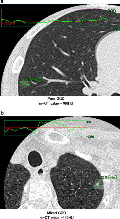
Methods: Between April 2001 and March 2010, a total of 44 nodules in 42 patients with pure or mixed GGOs ≤2 cm were retrospectively evaluated. Maximum diameter and one-dimensional mean CT (m-CT) value of the diameter were measured using a computer graphics support system (HOPE/DrABLE-EX, Fujitsu, Tokyo, Japan) that displays a CT density profile across the tumor.

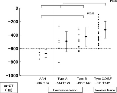
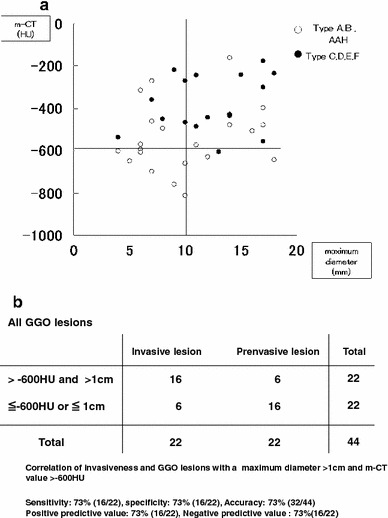
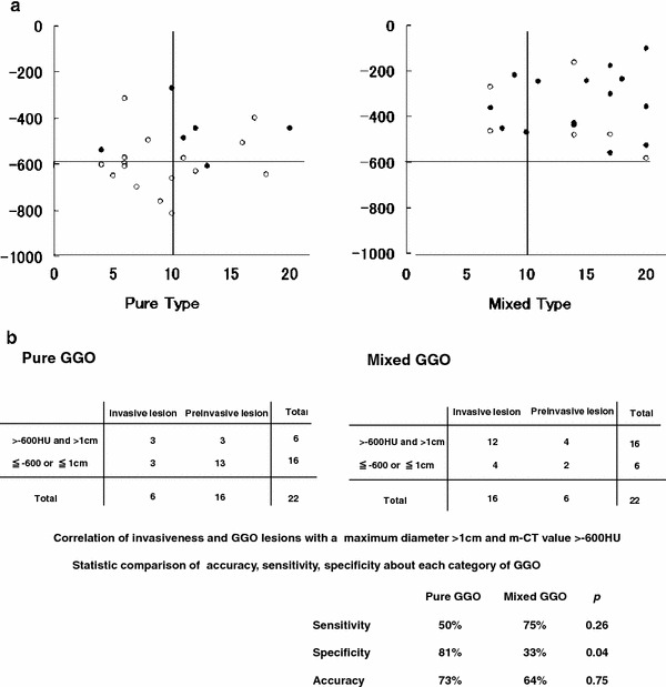
Results: m-CT values were -682 ± 64 HU (range) for AAH lesions, -544 ± 179 (range) for Type A lesions, -496 ± 147 (range) for Type B lesions, and -371 ± 142 (range) for invasive lesions. AAH lesions had a significantly lower m-CT value than Type B lesions. AAH, Type A, and Type B lesions had significantly lower m-CT values than invasive lesions (p < 0.05). All seven GGO lesions with a maximum diameter ≤1 cm and m-CT value ≤-600 HU were pre-invasive lesions, while 16 of 22 (73 %) cases with maximum diameter >1 cm and m-CT value >-600 HU were invasive lesions.

Conclusion: Observation may be indicated for GGO lesions with a maximum diameter ≤1 cm and m-CT value ≤-600 HU.


Figures

Fig. 1
Fig. 1
The CT image with a profile of the density across the tumor. The profile of pure GGO revealed an almost flat curve, and the m-CT value was calculated to be −760 HU (a). The profile of mixed GGO revealed that the CT value of the solid portion was higher than the GGO portion, and the m-CT value was calculated to be −440 HU (b)
Fig. 2
Fig. 2
The m-CT values were −682 ± 64 HU (range) for AAH lesions, −544 ± 179 (range) for Type A lesions, −496 ± 147 (range) for Type B lesions, and −371 ± 142 (range) for invasive lesions. AAH lesions had a significantly lower m-CT value than Type B lesions. AAH, Type A, and Type B lesions had a significantly lower m-CT values than invasive lesions (p < 0.05). No significant differences in m-CT values were observed between AAH and Type A lesions or between Type A and Type B lesions. The m-CT value of AAH lesions were all ≤−600 HU, while most of the m-CT values of invasive lesions were >−600 HU
Fig. 3
Fig. 3
a, b Relationship between m-CT value and maximum diameter of all GGO lesions. All seven GGO lesions with a maximum diameter ≤1 cm in the maximum diameter and m-CT value ≤−600 HU were pre-invasive lesions, while 16 of 22 (73 %) with a maximum diameter >1 cm and m-CT value >−600 HU in m-CT were invasive lesions
Fig. 4
Fig. 4
a, b Relationship between m-CT value and maximum diameter of pure and mixed type GGO lesions. GGO lesions ≤1 cm in maximum diameter and ≤−600 HU in m-CT value are all pure type. Although, GGO lesions of both categories with a maximum diameter >1 cm and m-CT value >−600 HU are highly diagnosed as invasive lesions, specificity of pure GGO were significantly better than that of mixed GGO

References

    1. Henschke CI, McCauley DI, Yankelevitz D, Naidich DP, McGuinness G, Miettinen OS, et al. Early lung cancer action project: overall design and findings from baseline screening. Lancet. 1999;354:99–105. doi: 10.1016/S0140-6736(99)06093-6. - DOI - PubMed
    1. Parkin DM, Moss SM. Lung cancer screening. Improved survival but no reduction in deaths—the role of “overdiagnosis”. Cancer. 2000;89:2369–2376. doi: 10.1002/1097-0142(20001201)89:11+<2369::AID-CNCR10>3.0.CO;2-A. - DOI - PubMed
    1. Patz EF, Goodman PC, Bepler G. Screening for lung cancer. N Engl J Med. 2000;343:1627–1633. doi: 10.1056/NEJM200011303432208. - DOI - PubMed
    1. Travis WD, Colby TV, Corrin B, Shimosato Y, Brambilla E. Histological typing of lung and pleural tumors. In: Sobin LH, editor. World Health Organization international histological classification of tumors. 3. New York: Springer; 1999. pp. 58–63.
    1. Noguchi M, Morikawa A, Kawasaki M, Matsuno Y, Yamada T, Hirohashi S, et al. Small adenocarcinoma of the lung. Histologic characteristics and prognosis. Cancer. 1995;75:2844–2852. doi: 10.1002/1097-0142(19950615)75:12<2844::AID-CNCR2820751209>3.0.CO;2-#. - DOI - PubMed

MeSH terms