Review
. 2012 Jun 29;13(7):608-18.
doi: 10.1038/embor.2012.73.

The base of the cilium: roles for transition fibres and the transition zone in ciliary formation, maintenance and compartmentalization

Affiliations
Review

The base of the cilium: roles for transition fibres and the transition zone in ciliary formation, maintenance and compartmentalization

Jeremy F Reiter et al. EMBO Rep. .

Abstract

Both the basal body and the microtubule-based axoneme it nucleates have evolutionarily conserved subdomains crucial for cilium biogenesis, function and maintenance. Here, we focus on two conspicuous but underappreciated regions of these structures that make membrane connections. One is the basal body distal end, which includes transition fibres of largely undefined composition that link to the base of the ciliary membrane. Transition fibres seem to serve as docking sites for intraflagellar transport particles, which move proteins within the ciliary compartment and are required for cilium biogenesis and sustained function. The other is the proximal-most region of the axoneme, termed the transition zone, which is characterized by Y-shaped linkers that span from the axoneme to the ciliary necklace on the membrane surface. The transition zone comprises a growing number of ciliopathy proteins that function as modular components of a ciliary gate. This gate, which forms early during ciliogenesis, might function in part by regulating intraflagellar transport. Together with a recently described septin ring diffusion barrier at the ciliary base, the transition fibres and transition zone deserve attention for their varied roles in forming functional ciliary compartments.


Conflict of interest statement

The authors declare that they have no conflict of interest.

Figures
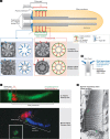
Figure 1
Figure 1
The transition fibres, and the transition zone with its associated ciliary necklace, represent evolutionarily conserved features of the basal body–ciliary organelle. (A) All cilia have a microtubule-based axoneme that emerges from a centriolar structure termed the ‘basal body’. The TFs and TZ are depicted schematically together with representative TEMs from a Caenorhabditis elegans sensory cilium and human oviduct primary cilium. Y-link structures organize, or make up, the ciliary necklace present on the ciliary surface (shown as beads). In nematodes, the basal body almost completely degenerates after ciliogenesis, retaining only TFs. The nature and function of the inner singlet microtubules seen in the TEM cross-sections of C. elegans cilia are unknown. The ciliary compartment is highlighted in yellow. (B) Substructures of the centriolar barrel, with the reported localization of several components shown. (C) Fluorescence microscopy images of markers showing the basal bodies, TZs and axonemes of C. elegans and mammalian cilia. The two C. elegans cilia show an IFT protein (DYF-11; green) marking the basal body (TF region) and axoneme, and MKS-5 (red) marking the TZ. The mouse cilium shows the basal body (γ-tubulin; red), TZ (membrane-associated TMEM231 forming a ring; green) and the axoneme (acetylated tubulin; blue). (D) Freeze–fracture scanning electron micrograph of a hamster respiratory cilium, showing evidence that the ciliary necklace is in fact a spiral, with an approximate 8° angle of pitch with respect to the ciliary axis (arrows show apparent start and end points of the bead-like particles on the membrane). C. elegans images modified with permission from [5]; oviduct TEM from [55]; TMEM231 data from [36]; ciliary necklace from [24]. IFT, intraflagellar transport; MT, microtubule; TEM, transmission electron migrograph; TF, transition fibre; TZ, transition zone.
Figure 2
Figure 2
Roles of the basal body distal end and transition zone region during two phases of primary cilium formation (‘early’ and ‘late’). In many mammalian cell types, the first ciliogenic event involves the binding of a CV to the distal end of the mother centriole, probably through distal appendages (1). A (presumably immature) TZ region begins to emerge and invaginate the CV, the membrane surface of which grows through fusion of secondary vesicles (2–3). The basal body-CV can migrate to the plasma membrane (3) and then fuse with it (4), at which point the maturing TZ forms the ciliary gate (5). Complete formation of the axoneme and functional cilium is an IFT/BBS protein-dependent process (6); IFT/BBS proteins present on the undocked basal body might perform specific transport roles or might simply be trafficked for eventual assembly as functional IFT particles. A different pathway (7) followed by other cells types might not involve a CV, but rather, have the basal body docking directly with the membrane; a TZ might start to form just before, during and after this step. Also, basal body migration does not occur in all cell types. CV, ciliary vesicle; IFT, intraflagellar transport; BBS, Bardet–Biedl syndrome; TZ, transition zone.
Figure 3
Figure 3
Regulation of membrane diffusion and transport by ciliary gate structures present in the TF–TZ region. Vesicles bearing ciliary membrane proteins dock at the base of cilia in proximity to the TF physical barrier. Ciliary cargo might be transported into cilia by IFT particles (shown moving bidirectionally along the axoneme), which dock at the TFs and functionally interact with TZ proteins. Shown below are fluorescence images of SEPT2 (green) at the base of a mammalian cilium (marked by acetylated tubulin; red), as well as of the tubulin quality control protein RP2 (green) within the basal body (TF region; marked by IFT protein in red) of C. elegans cilia. A RanGDP-RanGTP gradient across the cytosol and ciliary compartment is also depicted. Refer to text for further details. SEPT2 images modified with permission from [62]; original finding of RP2 ring at base of cilia published in [61]. RP2, retinitis pigmentosa 2; SEPT2, septin 2; TF, transition fibre; TZ, transition zone.
None
Michel R Leroux
None
Oliver E Blacque
None
Jeremy F Reiter

References

    1. Goetz SC, Anderson KV (2010) The primary cilium: a signalling centre during vertebrate development. Nat Rev Genet 11: 331–344 - PMC - PubMed
    1. Baker K, Beales PL (2009) Making sense of cilia in disease: the human ciliopathies. Am J Med Genet C Semin Med Genet 151C: 281–295 - PubMed
    1. Hildebrandt F, Benzing T, Katsanis N (2011) Ciliopathies. N Engl J Med 364: 1533–1543 - PMC - PubMed
    1. Deane JA, Cole DG, Seeley ES, Diener DR, Rosenbaum JL (2001) Localization of intraflagellar transport protein IFT52 identifies basal body transitional fibers as the docking site for IFT particles. Curr Biol 11: 1586–1590 - PubMed
    1. Williams CL et al. (2011) MKS and NPHP modules cooperate to establish basal body/transition zone membrane associations and ciliary gate function during ciliogenesis. J Cell Biol 192: 1023–1041 - PMC - PubMed

Publication types

Substances