. 2012 Jul 13;337(6091):189-94.
doi: 10.1126/science.1222804. Epub 2012 May 31.

Crystal structure of the heterodimeric CLOCK:BMAL1 transcriptional activator complex

Affiliations

Crystal structure of the heterodimeric CLOCK:BMAL1 transcriptional activator complex

Nian Huang et al. Science. .

Abstract

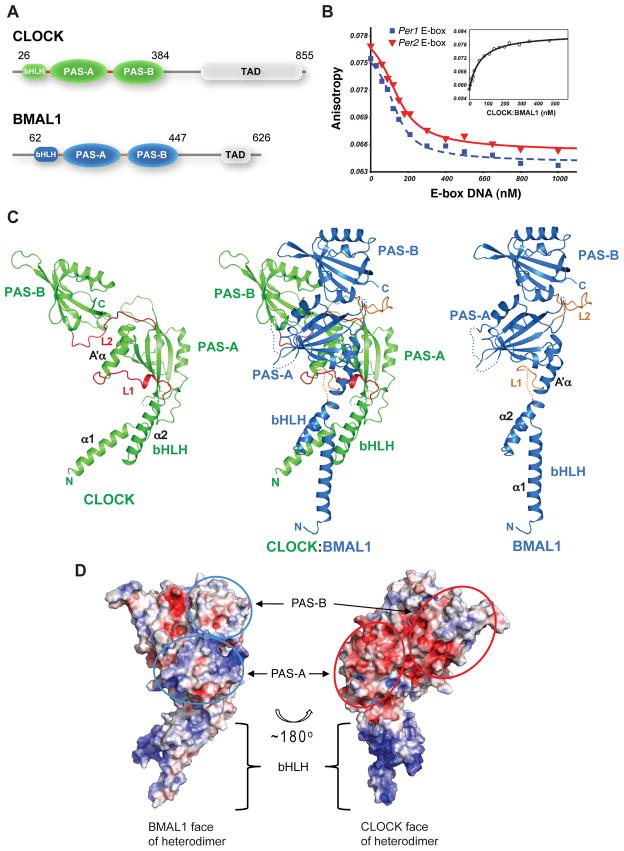
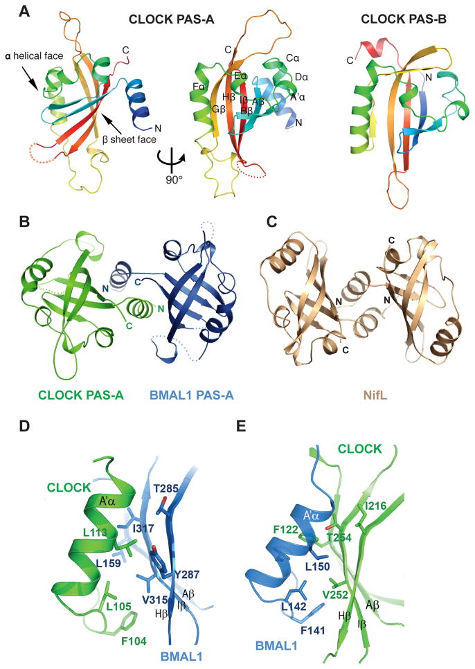
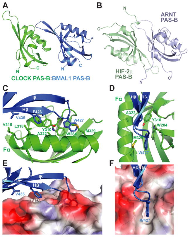
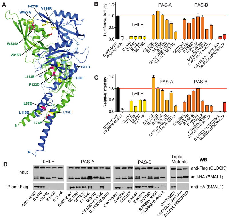
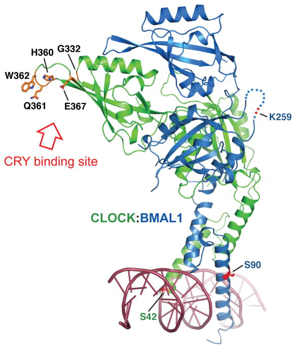
The circadian clock in mammals is driven by an autoregulatory transcriptional feedback mechanism that takes approximately 24 hours to complete. A key component of this mechanism is a heterodimeric transcriptional activator consisting of two basic helix-loop-helix PER-ARNT-SIM (bHLH-PAS) domain protein subunits, CLOCK and BMAL1. Here, we report the crystal structure of a complex containing the mouse CLOCK:BMAL1 bHLH-PAS domains at 2.3 Å resolution. The structure reveals an unusual asymmetric heterodimer with the three domains in each of the two subunits--bHLH, PAS-A, and PAS-B--tightly intertwined and involved in dimerization interactions, resulting in three distinct protein interfaces. Mutations that perturb the observed heterodimer interfaces affect the stability and activity of the CLOCK:BMAL1 complex as well as the periodicity of the circadian oscillator. The structure of the CLOCK:BMAL1 complex is a starting point for understanding at an atomic level the mechanism driving the mammalian circadian clock.


Figures

Fig. 1
Fig. 1
Overall structure of mouse CLOCK:BMAL1. (A) Domain organization of CLOCK and BMAL1. Crystals were obtained from the truncated proteins (indicated by the amino acid residue number) encompassing the bHLH-PAS-AB domains. (B) DNA-binding affinity of the truncated CLOCK:BMAL1 complex measured by fluorescence anisotropy. Dissociation constant (Kd) of the fluorescein labeled mPer2 E2-box DNA was 59 ± 7.3 nM by direct binding to CLOCK:BMAL1 (inset). Using unlabeled DNA probes as competitor, the Kd’s of unlabeled 18-mer mPer1 E1-box DNA (blue) and mPer2 E2-box DNA (red) (40) were 9.0 ± 2.3 nM and 13 ± 2.0 nM, respectively. (See Materials and Method for details). (C) Ribbon diagram of CLOCK:BMAL1 heterodimer (center). The CLOCK subunit is colored green, BMAL1 blue. Each individual domain is labeled. The CLOCK (left) and BMAL1 (right) subunits are also shown separately to illustrate their different spatial domain arrangements. The linker regions between domains in the two subunits (L1 and L2) are highlighted in red or orange color. Flexible loops lacking density are indicated by dotted lines. (D) Electrostatic potentials of CLOCK:BMAL1 heterodimer showing that the surfaces composed of CLOCK PAS domains (red ovals, right) have mostly negative potentials while the surfaces of BMAL1 PAS domains (blue ovals, left) are mostly positive or neutral. The colors are ramped from negative potential −5 kT/q (red) to positive 5 kT/q (blue).
Fig. 2
Fig. 2
Structure and interaction of the PAS-A domains of CLOCK:BMAL1. (A) Ribbon representations of CLOCK PAS-A domain. Secondary structures are color ramped from blue to red and labeled from the A′α helix located N-terminal to the canonical PAS domain fold, in an alphabetical progression through the whole domain.. The CLOCK PAS-B domain is also shown for comparison. (B) Dimerization of the two PAS-A domains in CLOCK:BMAL1, looking down the approximate two-fold symmetry axis. (C) Similar domain-swapped structure of the redox sensing PAS domain of NifL from A. vinelandii (pdb: 2GJ3). (D) Left panel, detailed interface between A′α helix of CLOCK PAS-A (green) and the β sheet face of BMAL1 PAS-A (blue). Right panel, the corresponding interface between A′α helix of BMAL1 PAS-A and the β sheet face of CLOCK PAS-A.
Fig. 3
Fig. 3
Interface between CLOCK:BMAL1 PAS-B domains. (A) The spatial arrangement of the two PAS-B domains in CLOCK:BMAL1. (B) Antiparallel orientation of β sheet-mediated interaction between isolated HIF-2α:ARNT PAS-B domains (pdb: 3F1P). (C) Detailed interface between CLOCK:BMAL1 PAS-B domains. (D) Front facing view of CLOCK:BMAL1 PAS-B interface highlighting role of BMAL1 Trp427and CLOCK Trp284 interaction. (E) Side view of PAS-B interface displaying surface electrostatic potential of CLOCK PAS-B. (F) Front facing view of CLOCK surface electrostatic potential displaying the binding pocket for BMAL1 Trp427. The color scheme used is the same as in Fig. 1D.
Fig. 4
Fig. 4
Functional analysis of CLOCK:BMAL1 mutants. (A) Locations of domain interface mutants in CLOCK (green) and BMAL1 (blue). (B) Per2 promoter:Luciferase reporter assays to evaluate the effects of structure-based mutations on transactivation by full-length CLOCK:BMAL1. Data are average of 2 independent experiments performed in duplicate. (C) Bimolecular fluorescence complementation (BiFC) experiments on the same set of mutants in CLOCK:BMAL1 truncated constructs. The fluorescent intensities of WT and mutant CLOCK:BMAL1 bHLH-PAS-AB constructs (for details, see Supplementary Materials and Methods) were quantified using data from 3 independent experiments. (E) Coimmunoprecipitation experiments assessing the association of CLOCK and BMAL1 in full-length WT and mutant proteins. Anti-FLAG affinity gel was used to precipitate FLAG tagged CLOCK along with the tightly associated BMAL1, which is HA tagged. The Western blots using an anti-HA antibody were then performed to detect the association of WT and mutant BMAL1 with CLOCK constructs. The Co-IP data are representative of at least 3 independent replicates, with the exception of C:W284A which had stronger co-IP interaction in other experiments, but on average was weaker than WT CLOCK.
Fig. 5
Fig. 5
Mutations that reduce repression of CLOCK:BMAL1 transactivation by CRY localize to CLOCK PAS-B HI loop. CRY-derepressing mutations arising from a random mutagenesis screen: G332E, H360Y, E367K (18) or directed mutagenesis study, Q361P/W362R (19) are predominantly found on the β-sheet face of CLOCK PAS-B domain and are fully solvent accessible. Residues mutated in these studies are in orange. The locations of the SUMOylation site on BMAL1 PAS-A (K259) (41), the Casein kinase 2 phosphorylation site on BMAL1 (S90) (42), and the phosphorylation site on CLOCK (S42) (43) are also indicated. A double strand DNA is modeled based on the superposition with USF-DNA complex structure (25).

Comment in

References

    1. King DP, et al. Cell. 1997;89:641. - PMC - PubMed
    1. Antoch MP, et al. Cell. 1997;89:655. - PMC - PubMed
    1. Reppert SM, Weaver DR. Nature. 2002;418:935. - PubMed
    1. Lowrey PL, Takahashi JS. Annu Rev Genom Hum Genet. 2004;5:407. - PMC - PubMed
    1. Lowrey PL, Takahashi JS. Adv Genet. 2011;74:175. - PMC - PubMed

Publication types

MeSH terms

Associated data