. 2012 Dec;56(6):2163-71.
doi: 10.1002/hep.25871. Epub 2012 Sep 17.

JD induced pluripotent stem cell-derived hepatocytes faithfully recapitulate the pathophysiology of familial hypercholesterolemia

Affiliations

JD induced pluripotent stem cell-derived hepatocytes faithfully recapitulate the pathophysiology of familial hypercholesterolemia

Max A Cayo et al. Hepatology. 2012 Dec.

Abstract

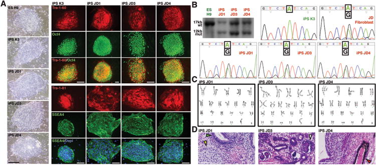
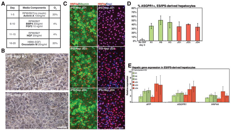
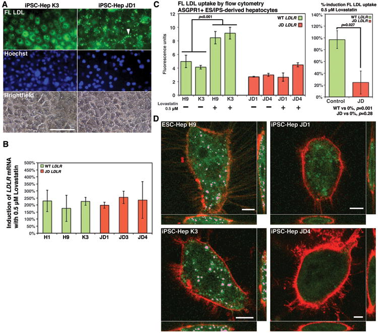
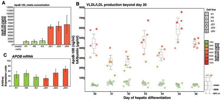
Elevated levels of low-density lipoprotein cholesterol (LDL-C) in plasma are a major contributor to cardiovascular disease, which is the leading cause of death worldwide. Genome-wide association studies (GWAS) have identified 95 loci that associate with control of lipid/cholesterol metabolism. Although GWAS results are highly provocative, direct analyses of the contribution of specific allelic variations in regulating LDL-C has been challenging due to the difficulty in accessing appropriate cells from affected patients. The primary cell type responsible for controlling cholesterol and lipid flux is the hepatocyte. Recently, we have shown that cells with hepatocyte characteristics can be generated from human induced pluripotent stem cells (iPSCs). This finding raises the possibility of using patient-specific iPSC-derived hepatocytes to study the functional contribution of GWAS loci in regulating lipid metabolism. To test the validity of this approach, we produced iPSCs from JD a patient with mutations in the low-density lipoprotein receptor (LDLR) gene that result in familial hypercholesterolemia (FH). We demonstrate that (1) hepatocytes can be efficiently generated from FH iPSCs; (2) in contrast to control cells, FH iPSC-derived hepatocytes are deficient in LDL-C uptake; (3) control but not FH iPSC-derived hepatocytes increase LDL uptake in response to lovastatin; and (4) FH iPSC-derived hepatocytes display a marked elevation in secretion of lipidated apolipoprotein B-100.

Conclusion: Cumulatively, these findings demonstrate that FH iPSC-derived hepatocytes recapitulate the complex pathophysiology of FH in culture. These results also establish that patient-specific iPSC-derived hepatocytes could be used to definitively determine the functional contribution of allelic variation in regulating lipid and cholesterol metabolism and could potentially provide a platform for the identification of novel treatments of cardiovascular disease. (HEPATOLOGY 2012).


Figures

Fig. 1
Fig. 1. Generation of induced pluripotent stem cells from ‘JD’ fibroblasts
(a) Micrographs showing colony morphology and expression of characteristic markers of pluripotency (Tra-1-60, OCT4, Tra-1-81, SSEA4) identified by immunocytochemistry in huESCs (H9), control hiPSC (K3), and three independently derived ‘JD’ iPSC lines (JD1, JD3, JD4). (b) Southern blot analysis (upper left panel) using a probe from LDLR exon 18 to identify a 17kb wild-type BamH1 fragment (wt) in control cells and an additional 12kb mutant fragment (mut) in ‘JD’ hiPSCs that represents the maternal allele. DNA sequencing revealed the presence of the paternal A-G transition exclusively in ‘JD’ fibroblasts and ‘JD’ iPSCs. (c) ‘JD’ hiPSCs retain a normal chromosomal arrangement as revealed by karyotyping. (d) Teratomas generated in immunocompromised mice from ‘JD’ hiPSCs contained cell types representative of all germ layers (arrowheads). Scale bars = 100μM.
Fig. 2
Fig. 2. Hepatocyte–like cells can be efficiently derived from ‘JD’ hiPSCs
(a) Schematic showing procedure used to generate hepatocytes from human pluripotent stem cells. (b) Micrographs showing morphology of hepatocytes derived from WT and ‘JD’ hiPSCs. (c) Immunocytochemistry revealing the presence of HNF4a in the nucleus (red) and Albumin in the cytoplasm (green) of ‘JD’–hiPSC–derived hepatocytes. Nuclei are identified by DAPI staining (blue). (d) Bar graph illustrating the efficiency and reproducibility (n=3 independent experiments) of generating ASGPR positive hepatocytes from control (H1, H9, K3) and ‘JD’ (JD1, JD3, JD4) pluripotent stem cells. No significant difference was observed between control and ‘JD’ cells (ANOVA, p>0.05). (f) Bar graph showing result of real time qRT-PCR analyses of characteristic hepatocyte mRNAs (AFP, ASGPR1, HNF4A) in undifferentiated pluripotent stem cells and in hepatocytes generated from control (H1, H9, K3) and ‘JD’ (JD1, JD3, JD4) pluripotent stem cells. No significant difference was observed between control and ‘JD’ cells (ANOVA, p>0.05). Scale bars = 100μM.
Fig. 3
Fig. 3. ‘JD’ hiPSC–derived hepatocytes exhibit deficiencies in uptake of LDL–C and in their response to lovastatin
(a) Micrographs showing localization of FL LDL (green) after incubation of control and ‘JD’ iPS cell–derived hepatocytes with BODIPY labeled LDL for 3.5 hours. Note clustering of FL LDL specifically on the surface of ‘JD’ cells (arrow). Staining with Hoechst dye identified nuclei (blue) and cell distribution is shown by bright field images. Scale bars = 100μM. (b) Bar graph shows percent increase in LDLR mRNA following lovastatin treatment of control (green bars) and ‘JD’ (red bars) hepatocytes. No statistically significance difference in the level of induction was observed between control and ‘JD’ cells (ANOVA p>0.05). (c) Bar graph (left panel) showing a significant (Student's t-test p<0.05) increase in FL LDL uptake was observed in response to lovastatin treatment by control hepatocytes (green bars) but not by ‘JD’ hepatocytes (red bars). Right bar graph shows that the differential response to lovastatin treatment between control (green bar) and ‘JD’ (red bar) hepatocytes is significant (Student's t-test, p<0.05). (d) Volume render images obtained by confocal microscopy showing that uptake of FL LDL (pink) could be identified in endosomes of lovastatin–treated control (ES-Hep H9, iPS-Hep K3) hepatocytes but not in ‘JD’ hepatocytes. Plasma membranes were identified using FM® 4-64 (Molecular Probes) (red). Scale bars = 20μM.
Fig. 4
Fig. 4. ApoB-100 secretion is elevated in ‘JD’ iPSC-derived hepatocytes
(a) Bar graphs showing concentration of lipidated ApoB secreted into the medium by control (H1, H9, and K3) (green boxes) or ‘JD’ (JD1, JD3, JD4) (red boxes) iPSC-derived hepatocytes as determined by ELISA (n=3 independent differentiations). No significant difference was observed within control or mutant groups; however, secreted ApoB-levels were significantly elevated in ‘JD’ compared to control hepatocytes (p<0.05 Tukey-Kramer post hoc multiple comparison test). (b) Box and whisker plot showing that elevated ApoB production is maintained in hepatocytes derived from ‘JD’ iPSCs compared to control hepatocytes after extended culture (days 20 – 26) of the differentiated cells. (c) Bar graph showing level of APOB mRNA in hepatocytes derived from control (H1, H9, and K3) (green boxes) or ‘JD’ (JD1, JD3, JD4) (red boxes) pluripotent stem cells as determined by qRT-PCR analyses. APOB mRNA levels were not significantly different between cell lines (ANOVA, p>0.05).

References

    1. Rosamond W, Flegal K, Friday G, et al. Heart disease and stroke statistics--2007 update: a report from the American Heart Association Statistics Committee and Stroke Statistics Subcommittee. Circulation. 2007;115:e69–171. - PubMed
    1. Baigent C, Blackwell L, Emberson J, et al. Efficacy and safety of more intensive lowering of LDL cholesterol: a meta-analysis of data from 170,000 participants in 26 randomised trials. Lancet. 2010;376:1670–1681. - PMC - PubMed
    1. Teslovich TM, Musunuru K, Smith AV, et al. Biological, clinical and population relevance of 95 loci for blood lipids. Nature. 2010;466:707–713. - PMC - PubMed
    1. Si-Tayeb K, Noto FK, Nagaoka M, et al. Highly efficient generation of human hepatocyte-like cells from induced pluripotent stem cells. Hepatology. 2010;51:297–305. - PMC - PubMed
    1. Sullivan GJ, Hay DC, Park IH, et al. Generation of functional human hepatic endoderm from human induced pluripotent stem cells. Hepatology. 2010;51:329–335. - PMC - PubMed

Publication types

MeSH terms