. 2012 Jul 13;287(29):24832-43.
doi: 10.1074/jbc.M112.370783. Epub 2012 May 31.

A regulatory feedback loop between Ca2+/calmodulin-dependent protein kinase kinase 2 (CaMKK2) and the androgen receptor in prostate cancer progression

Affiliations

A regulatory feedback loop between Ca2+/calmodulin-dependent protein kinase kinase 2 (CaMKK2) and the androgen receptor in prostate cancer progression

Loukia G Karacosta et al. J Biol Chem. .

Abstract

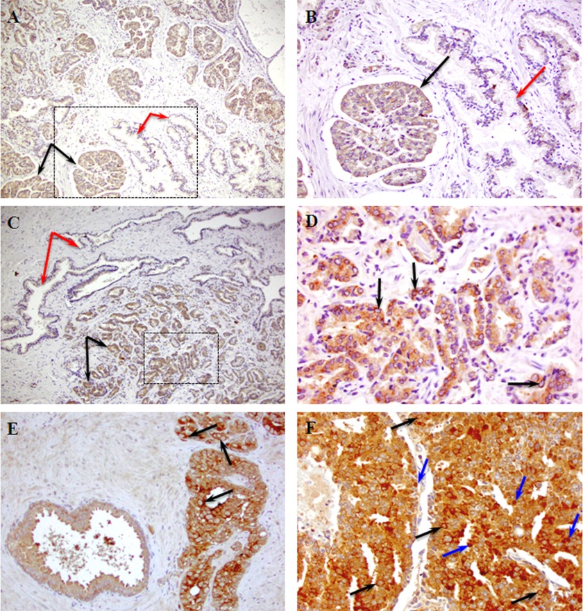
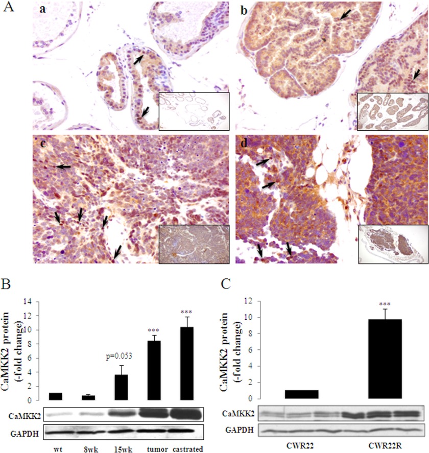
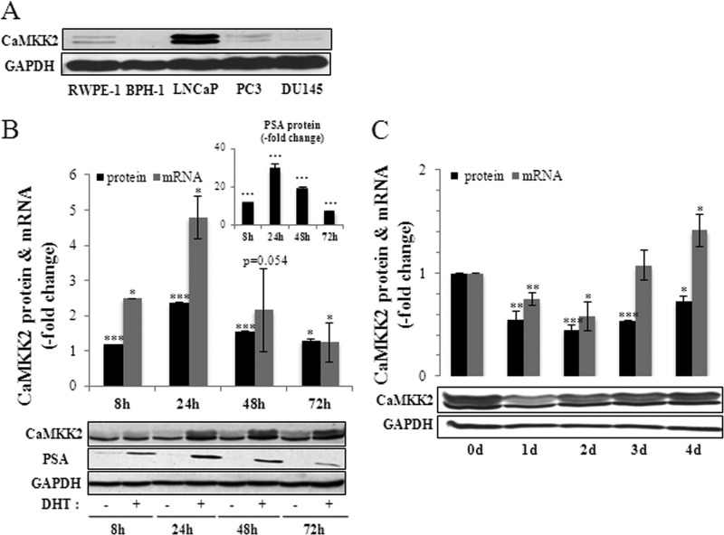
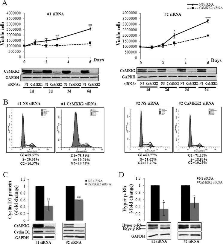
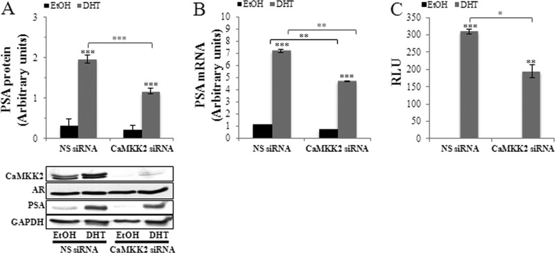
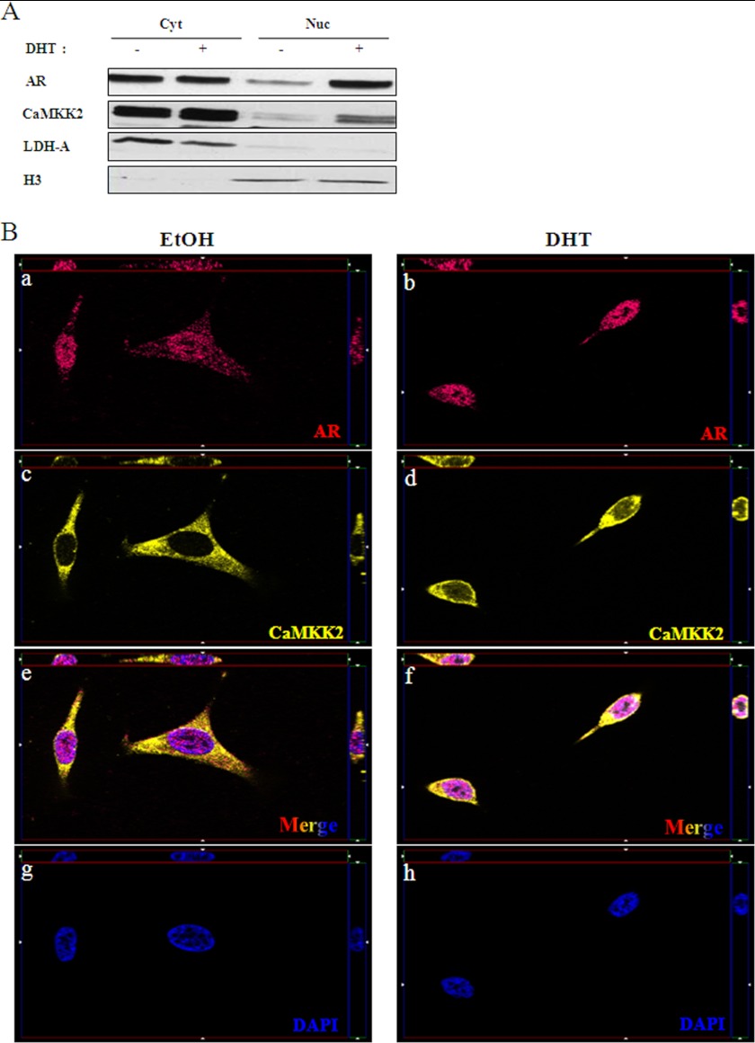
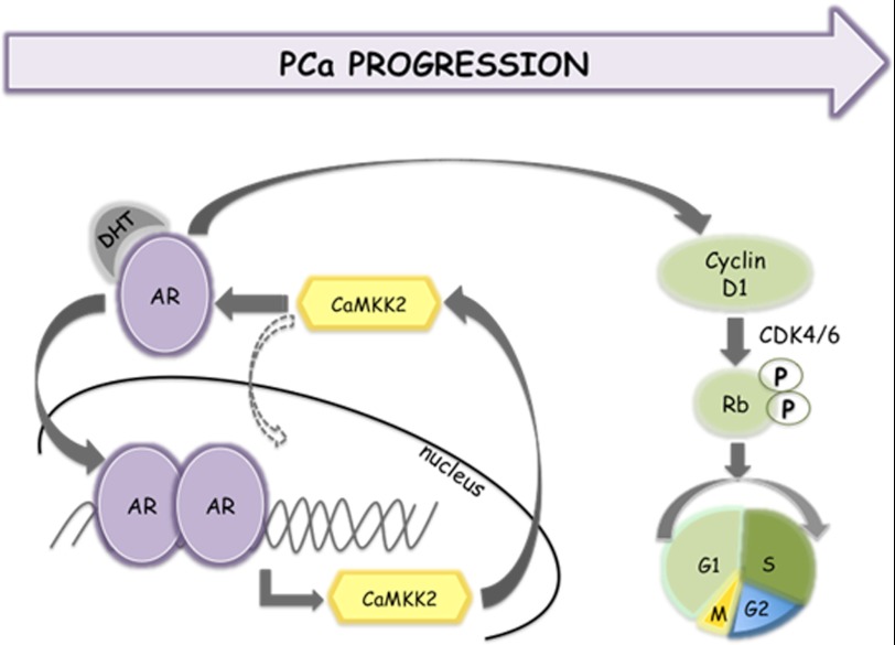
The androgen receptor (AR) plays a critical role in prostate cancer (PCa) progression, however, the molecular mechanisms by which the AR regulates cell proliferation in androgen-dependent and castration-resistant PCa are incompletely understood. We report that Ca(2+)/calmodulin-dependent kinase kinase 2 (CaMKK2) expression increases and becomes nuclear or perinuclear in advanced PCa. In the TRAMP (transgenic adenocarcinoma of mouse prostate) model of PCa, CaMKK2 expression increases with PCa progression with many cells exhibiting nuclear staining. CaMKK2 expression is higher in human castration-resistant tumor xenografts compared with androgen-responsive xenografts and is markedly higher in the AR-expressing, tumorigenic cell line LNCaP compared with cell lines that are AR-nonexpressing and/or nontumorigenic. In LNCaP cells, dihydrotestosterone induced CaMKK2 mRNA and protein expression and translocation of CaMKK2 to the nucleus. Conversely, androgen withdrawal suppressed CaMKK2 expression. Knockdown of CaMKK2 expression by RNAi reduced LNCaP cell proliferation and increased percentages of cells in G(1) phase, whereas correspondingly reducing percentages in S phase, of the cell cycle. CaMKK2 knockdown reduced expression of the AR target gene prostate-specific antigen at both mRNA and protein levels, AR transcriptional activity driven by androgen responsive elements from the prostate-specific probasin gene promoter and levels of the AR-regulated cell cycle proteins, cyclin D1 and hyperphosphorylated Rb. Our results suggest that in PCa progression, CaMKK2 and the AR are in a feedback loop in which CaMKK2 is induced by the AR to maintain AR activity, AR-dependent cell cycle control, and continued cell proliferation.


Figures

FIGURE 1.
FIGURE 1.
CaMKK2 expression in clinical specimens of PCa. Shown are representative images of CaMKK2 IHC staining in patient samples with various histological Gleason scores3 representing disease advancement and predicting aggressiveness of prostate tumors. A, CaMKK2 shows stronger epithelial staining in malignant glands (black arrows) compared with adjacent benign glands (red arrows) in a patient specimen of GS 3 + 4. B, image A (boxed area) at higher magnification. C, CaMKK2 expression differences in a specimen of GS 3 + 4 from a second patient. D, image C (malignant area, boxed) at higher magnification. Note the perinuclear CaMKK2 staining (arrows). E, CaMKK2 expression is higher in a GS 4 + 4 tumor area (right) compared with an adjacent HGPIN area (left). Arrows indicate perinuclear CaMKK2 staining. F, CaMKK2 staining is uniformly intense in a GS 4 + 5 tumor specimen. Note the appearance of nuclear CaMKK2 staining (blue arrows) apart from the perinuclear staining (black arrows). Magnifications: A and C, ×10; B and E, ×20; D and F, ×40.
FIGURE 2.
FIGURE 2.
Expression of CaMKK2 during tumor progression in transgenic and xenograft models of PCa. A, shown are representative images of CaMKK2 IHC staining in the TRAMP mouse over a course of PCa progression. a, nontransgenic (C57BL/6 x FVB) prostate tissue. b, prostate tissue of a 15-week-old TRAMP mouse. c, prostate tumor of a 27-week-old TRAMP mouse. d, prostate tumor of a 24-week-old castrated TRAMP mouse. Arrows indicate nuclear CaMKK2 staining. Magnifications: a-d, ×40; a-d, insets, ×10. B, samples were harvested at the indicated times of tumor progression (as in panel A with the addition of prostate tissue from an 8 weeks TRAMP mouse) and assessed by Western blotting for CaMKK2 protein expression. GAPDH was used as loading control. The graph represents mean ± S.E. (n = 5), ***, p < 0.001, relative to wt CaMKK2 levels. C, CaMKK2 protein expression in human CWR22 and CWR22R tumor xenografts. Tumors were harvested from three independent CWR22 and CWR22R tumor-bearing mice and lysates were subjected to Western blotting for CaMKK2 protein expression. GAPDH was used as loading control. The graph represents mean ± S.E. (n = 3) normalized to CWR22 CaMKK2 protein levels. ***, p < 0.001, relative to CWR22.
FIGURE 3.
FIGURE 3.
Androgen regulation of CaMKK2 expression. A, relative levels of CaMKK2 protein in prostatic cell lines as assessed by Western blotting. GAPDH is used as loading control. B, DHT stimulates CaMKK2 expression. LNCaP cells were cultured in steroid-depleted media for 24 h then treated with 10 nm DHT or vehicle (EtOH) for the indicated time periods. Levels of CaMKK2 protein and mRNA, quantified by Western blotting and qRT-PCR, respectively, are shown with a representative blot below. Values are normalized to time-matched vehicle controls and represent mean ± S.E. (n = 3 independent experiments). *, p < 0.05; **, p < 0.01; ***, p < 0.001, relative to time-matched vehicle controls. PSA protein expression in response to 10 nm DHT is shown in the inset. GAPDH is used as loading control. C, steroid depletion reduces CaMKK2 expression. LNCaP cells were cultured in normal media for 72 h then in steroid-depleted media for the indicated time periods. Levels of CaMKK2 protein and mRNA, quantified by Western blotting and qRT-PCR, respectively, are shown with a representative blot below. Results represent mean ± S.E. (n = 3 independent experiments). *, p < 0.05; **, p < 0.01; ***, p < 0.001, relative to 0 day controls. GAPDH is used as loading control.
FIGURE 4.
FIGURE 4.
CaMKK2 regulation of PCa cell proliferation and cell cycle progression. A, CaMKK2 knockdown inhibits growth of LNCaP cells. LNCaP cells were transfected with either nonspecific (NS)- or CaMKK2-siRNAs and grown for the indicated time periods. Numbers of viable cells, determined by cell counting with trypan blue exclusion are shown with representative Western blots below. Results represent mean ± S.E. (n = 3 independent experiments). *, p < 0.05; **, p < 0.01; ***, p < 0.001, relative to NS siRNA time-matched controls. GAPDH is used as loading control. B, CaMKK2 knockdown arrests LNCaP cells in the G1 phase of the cell cycle. LNCaP cells were transfected with either NS- or CaMKK2-siRNAs, grown for 4 days, and analyzed by PI flow cytometry. Representative DNA content histograms (for each siRNA used) are shown. Results from this experiment and an additional independent experiment are averaged and shown in supplemental Fig. S5B. C and D, CaMKK2 knockdown reduces cyclin D1 and hyper p-Rb expression. LNCaP cells were transfected with either NS- or CaMKK2-siRNAs and grown for 72 h. Levels of cyclin D1 (C) and hyper p-Rb (D) proteins, quantified by Western blotting are shown with representative blots below. Results represent mean ± S.E. (n = 3 independent experiments). *, p < 0.05; **, p < 0.01, relative to NS-treated control cells. GAPDH is used as loading control.
FIGURE 5.
FIGURE 5.
CaMKK2 regulates transcriptional activity of the AR. A, CaMKK2 knockdown inhibits androgen-induced PSA protein expression. LNCaP cells were transfected with NS- or CaMKK2- siRNA (#2) in normal media for 48 h, switched to steroid-depleted media for 24 h, and then treated with 10 nm DHT or vehicle (EtOH) for an additional 24 h. Levels of PSA protein were quantified by Western blotting and normalized to GAPDH. A representative blot is shown below. GAPDH is used as loading control. Results represent mean ± S.E. in arbitrary units (n = 3 independent experiments). Note that AR protein levels are unaltered under these conditions. B, CaMKK2 knockdown inhibits androgen-induced PSA mRNA expression. LNCaP cells were transfected with NS- or CaMKK2-siRNA (#2) in normal media for 24 h, switched to steroid-depleted media for 72 h, and then treated with 10 nm DHT or vehicle (EtOH) for an additional 16 h. Levels of PSA mRNA were quantified by qRT-PCR and normalized to GAPDH. Results represent mean ± S.E. in arbitrary units (n = 3 independent experiments). C, CaMKK2 knockdown inhibits AR transcriptional activity. LNCaP cells were transfected with the probasin ARE luciferase reporter along with NS- or CaMKK2-siRNAs for 6 h, after which cells were switched to steroid-depleted media for 24 h then stimulated with either DHT (10 nm) or EtOH for an additional 16 h. Firefly luciferase activity normalized to Renilla luciferase activity is shown. Results represent mean relative light units (RLU) ± S.E. (n = 2 independent experiments). A-C, *, p < 0.05; **, p < 0.01; ***, p < 0.001, either of DHT-treated cells relative to EtOH-treated cells (over bars) or of NS relative to CaMKK2-siRNA-treated cells (over brackets).
FIGURE 6.
FIGURE 6.
DHT induces CaMKK2 nuclear translocation. A, LNCaP cells were cultured in steroid-depleted media for 24 h, then treated with 10 nm DHT or vehicle (EtOH) for an additional 16 h. Cells were then fractionated into cytoplasmic and nuclear compartments and assessed for CaMKK2 and AR expression by Western blotting. Lactate dehydrogenase (LDH-A) and histone H3 were used for assessing purity of cytoplasmic and nuclear fractions, respectively. Shown is a representative blot of two independent experiments. B, representative images of immunofluorescent staining for AR (a, b, red), CaMKK2 (c, d, yellow), DAPI (g, h, blue), and merged images (e, f, tricolor) and in LNCaP cells treated with 10 nm DHT or EtOH for 16 h. Single plane apotome (x-y plane) and z-sections (bordering the x-y planes) confirm CaMKK2 presence in the nucleus. Total magnification, ×630. Images are representative of two independent experiments.
FIGURE 7.
FIGURE 7.
Schematic model for a CaMKK2-AR feedback loop in PCa progression. See text for details.

Similar articles

Cited by

References

    1. Colomer J., Means A. R. (2007) Physiological roles of the Ca2+/CaM-dependent protein kinase cascade in health and disease. Subcell. Biochem. 45, 169–214 - PubMed
    1. Tokumitsu H., Enslen H., Soderling T. R. (1995) Characterization of a Ca2+/calmodulin-dependent protein kinase cascade. J. Biol. Chem. 270, 19320–19324 - PubMed
    1. Edelman A. M., Mitchelhill K. I., Selbert M. A., Anderson K. A., Hook S. S., Stapleton D., Goldstein E. G., Means A. R., Kemp B. E. (1996) Multiple Ca2+/calmodulin-dependent protein kinase kinases from rat brain. J. Biol. Chem. 271, 10806–10910 - PubMed
    1. Kitani T., Okuno S., Fujisawa H. (1997) Molecular cloning of Ca2+/calmodulin-dependent protein kinase kinase β. J. Biochem. 122, 243–250 - PubMed
    1. Anderson K. A., Means R. L., Huang Q. H., Kemp B. E., Goldstein E. G., Selbert M. A., Edelman A. M., Fremeau R. T., Means A. R. (1998) Components of a calmodulin-dependent protein kinase cascade, molecular cloning, functional characterization and cellular localization of Ca2+/calmodulin-dependent protein kinase kinase β. J. Biol. Chem. 273, 31880–31889 - PubMed

Publication types

MeSH terms

Substances