. 2012 Jul 27;287(31):26200-12.
doi: 10.1074/jbc.M112.349811. Epub 2012 May 31.

Optimal intensity shock wave promotes the adhesion and migration of rat osteoblasts via integrin β1-mediated expression of phosphorylated focal adhesion kinase

Affiliations

Optimal intensity shock wave promotes the adhesion and migration of rat osteoblasts via integrin β1-mediated expression of phosphorylated focal adhesion kinase

Jian-kun Xu et al. J Biol Chem. .

Abstract

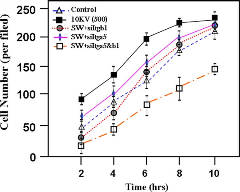
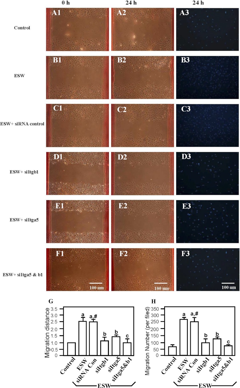
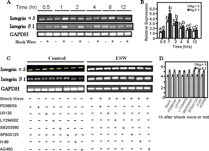
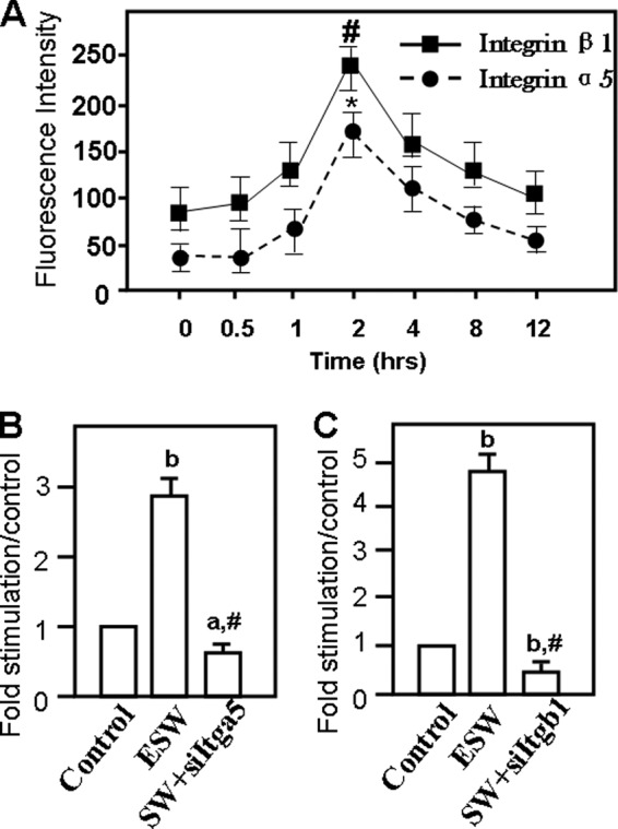
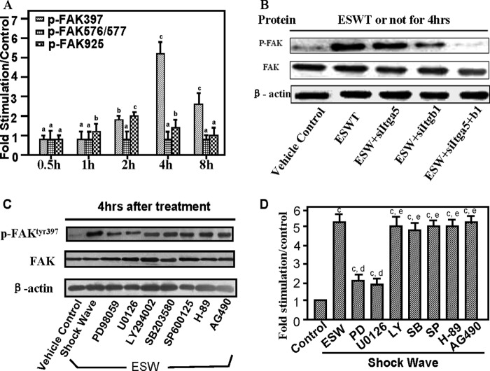
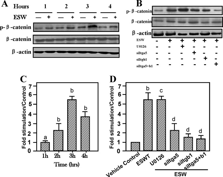
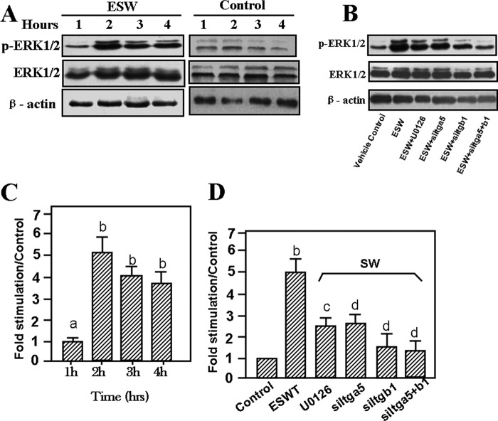
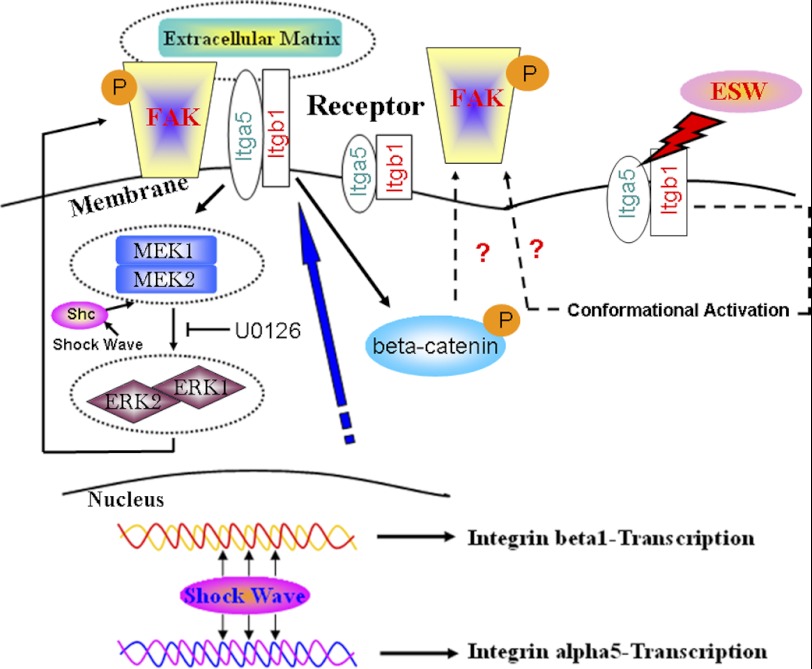
To search for factors promoting bone fracture repair, we investigated the effects of extracorporeal shock wave (ESW) on the adhesion, spreading, and migration of osteoblasts and its specific underlying cellular mechanisms. After a single period of stimulation by 10 kV (500 impulses) of shock wave (SW), the adhesion rate was increased as compared with the vehicle control. The data from both wound healing and transwell tests confirmed an acceleration in the migration of osteoblasts by SW treatment. RT-PCR, flow cytometry, and Western blotting showed that SW rapidly increased the surface expression of α5 and β1 subunit integrins, indicating that integrin β1 acted as an early signal for ESW-induced osteoblast adhesion and migration. It has also been found that a significant elevation occurred in the expression of phosphorylated β-catenin and focal adhesion kinase (FAK) at the site of tyrosine 397 in response to SW stimulation after the increasing expression of the integrin β1 molecule. When siRNAs of integrin α5 and β1 subunit were added, the level of FAK phosphorylation elevated by SW declined. Interestingly, the adhesion and migration of osteoblasts were decreased when these siRNA reagents as well as the ERK1/2 signaling pathway inhibitors, U0126 and PD98059, were present. Further studies demonstrated that U0126 could inhibit the downstream integrin-dependent signaling pathways, such as the FAK signaling pathway, whereas it had no influence on the synthesis of integrin β1 molecule. In conclusion, these data suggest that ESW promotes the adhesion and migration of osteoblasts via integrin β1-mediated expression of phosphorylated FAK at the Tyr-397 site; in addition, ERK1/2 are also important for osteoblast adhesion, spreading, migration, and integrin expression.


Figures

FIGURE 1.
FIGURE 1.
Optimal intensity of ESW (10 kV for 500 impulses) accelerated osteoblast adhesion. Data are presented as the mean ± S.D. (error bars) in triplicate independent experiments (n = 3). The data show that at the times of 2, 4, 6, 8, and 10 h after ESW treatment, the number of adhesive osteoblasts was significant higher than the number without ESW treatment. p < 0.01 as compared with the control group at the same period. When the siItgb1 was added prior to ESW treatment, the promotion of adhesion of osteoblasts by ESW was inhibited. p < 0.01 as compared with the ESW group at the same period. p > 0.05 as compared with the control group at the same period. It was observed that siItga5 also inhibited the ESW-induced adhesion although not as significantly as did siItgb1. The promotion of adhesion induced by ESW was abrogated, whereas integrin α5 and β1 subunits were silenced. p < 0.01 as compared with the ESW group at the same period.
FIGURE 2.
FIGURE 2.
ESW promoted migration of osteoblasts as shown in transwell tests and wound healing assays. The promotion could be inhibited by both siItgb1 and siItga5. Primary cultured osteoblasts were divided into six groups randomly; those were cells with (SW) (B1–B3) or without 10 kV for 500 impulses of ESWT (Control) (A1–A3) and with negative siRNA control (SW + siRNA control) (C1–C3), siItga5 (SW + siItga5) (E1–E3), siItgb1 (SW + siItgb1) (D1–D3), or both siItga5 and siItgb1 (F1–F3) for 6 h prior to ESWT. Results from the wound healing assays (G) and transwell tests (H) were consistent. Data are presented as the mean ± S.D. (error bars) (n = 6). a, p < 0.01; b, p < 0.05; c, p > 0.05 as compared with the control group. #, p > 0.05 as compared with the ESWT group. Scale bars, 100 μm.
FIGURE 3.
FIGURE 3.
A–D, ESW-induced elevations of mRNA level of α5 and β1 integrin of osteoblasts, peaking at 1 h (B). The specific inhibitors for signal transduction pathways had no influence on the integrin expression (C). A and C, representative electrophoretic images. The osteoblasts were harvested to extract total RNA 0.5, 1, 2, 4, 8, and 12 h after 500 impulses of 10-kV shock wave treatment. The cells without ESWT were run as control groups. After standardization of housekeeping gene expression, equal amounts of cDNA from each sample were subjected to 36 cycles to amplify Itga5 and Itgb1 mRNA expression. The values of the control group were normalized to 100%. a, p > 0.05; b, p < 0.01; c, p < 0.05 as compared with the control group at certain time periods. In addition, several signal transduction pathway inhibitors were added to the samples for 1 h prior to ESWT. 2 h after ESWT, the samples were collected to extract RNA and to analyze whether the Itga5 and Itgb1 mRNA were influenced by signal pathway inhibitors listed above. Our data showed that no influence on the expression of Itga5 or Itgb1 mRNA was observed under the conditions with or without inhibitors (p > 0.05). Error bars, S.D.
FIGURE 4.
FIGURE 4.
ESW enhanced integrin α5 and β1 subunit protein production in 2 h according to the data from flow cytometry analysis and Western blotting. For flow cytometry, osteoblasts from experimental groups and the control group were stained with PE-conjugated anti-rat Itga5 or Itgb1 antibody under the guidance of the manufacturer. It is shown that both Itga5 and Itgb1 proteins increased significantly in 2 h in the experimental group (A). *, p = 0.021; #, p < 0.01 as compared with the blank control group. Samples with or without ESWT and those associated with siItga5 and/or siItgb1 prior to ESW were subjected to radioimmune precipitation assay lysis. Western blotting was applied to analyze whether Itga5 and Itgb1 expression levels changed in the protein extractions. The same results are shown as those indicated by flow cytometry (B and C). In addition, the data from the ESW plus siRNA groups indicated that the siRNA reagents for both Itga5 and Itgb1 were effective (B and C). a, p < 0.05; b, p < 0.01 as compared with the blank control group. #, p < 0.01 as compared with ESW group. Results are presented with mean values ± S.E. (error bars) calculated from four paired triplicate experiments.
FIGURE 5.
FIGURE 5.
Evaluations of phosphorylation levels of focal adhesion kinase surrounding Tyr-397, Tyr-576/577, and Tyr-925. After experimental groups were subjected to direct exposure to 10 kV for 500 impulses of ESWT, we collected the extracts at 0.5, 1, 2, 4, or 8 h. Samples without ESWT were set as the control group. Then the extracts were quantified in triplicate using Western blotting and normalized by β-actin expression (A). A marked elevation of FAK phosphorylation (p-FAK) at Tyr-397 peaking at 4 h was observed, and we also observed a slight increase of FAK phosphorylation at Tyr-925 in 2 h after ESWT. ESW had no influence on the expression of FAK phosphorylation at Tyr-576/577 (Fig. 5A). A representative electrophoretic image of the study on FAK phosphorylation at Tyr-397 influenced by siRNAs of integrins is also depicted (B). 4 h after the optimal dose of ESWT, a decline in the expression of phosphorylated FAK at Tyr-397 was observed in the group with siRNA pretreatment. However, total protein expression levels of FAK were not affected by silencing of Itga5 and/or Itgb1 (Fig. 5B). Moreover, several specific cell signal pathway inhibitors, namely PD98059, U0126, LY294002, SB203580, SP600125, H-89, and AG490, were also added to the osteoblasts for 1 h, respectively, before ESWT. Both bands of total FAK and β-actin showed equal amounts of proteins subjected to protein electrophoresis (C). The data indicated that both PD98059 and U0126, unlike other inhibitors listed, inhibited ESW-induced FAK phosphorylation at Tyr-397 (D). Data represent the mean ± S.E. (error bars) in triplicate independent experiments (n = 3). The values of the control group were normalized to 100%. a, p > 0.05; b, p < 0.05; c, p < 0.01 as compared with the control group at the same time. d, p < 0.01; e, p > 0.05 versus ESW group.
FIGURE 6.
FIGURE 6.
Enhancement of β-catenin activity by ESW (500 impulses at 10 kV) stimulation after elevation of integrin α5 and β1 expression. ESW raised β-catenin phosphorylation in 3 h (A). ERK1/2 inhibitor U0126 did not alter the activation of β-catenin (B). Cytosolic extracts of osteoblasts treated with ESW in the presence of U0126 (with a final concentration of 20 μm) for 60 min prior to ESW were subjected to Western blotting. Phosphorylated β-catenin and β-catenin were probed with anti-phospho-β-catenin and β-catenin primary monoclonal antibodies, respectively. C, note that in comparison with the control, ESW exposure markedly elevated the activation of β-catenin. Further studies on the relationship between expression of integrins and β-catenin by transfection have shown that knocking out integrins led to base-line level expression of activation of β-catenin (B and D). D, summary of the results (mean ± S.E. (error bars), n = 4, triplicate in each experiment). a, p > 0.05 as compared with the control at the same period. b, p < 0.01 as compared with the control at the same period. c, p > 0.05 as compared with the ESW group. d, p < 0.01 as compared with the ESW group.
FIGURE 7.
FIGURE 7.
ESW activated ERK phosphorylation after treatment. ERK1/2 phosphorylation was increased in 2 h (A and C). The higher production of phosphorylated ERK1/2 persisted for 4 h (A and C). Osteoblasts were serum-deprived for 12 h before treatment with 10 kV for 500 impulses in the absence or presence of U0126 and siRNAs for the indicated time. Western blotting analysis was performed with antibodies against ERK and its phosphorylated forms (p-ERK) (B). C and D, summary of the results (mean ± S.E. (error bars), n = 4, triplicate in each experiment). a, p > 0.05 as compared with the control at the same period. b, p < 0.01 as compared with the control at the same period. c, p > 0.05 as compared with the control group. d, p < 0.01 as compared with the ESW group.
FIGURE 8.
FIGURE 8.
Hypothetical model elucidating the regulation of phosphorylated FAK expression through an integrin α5 and β1-mediated MEK-ERK1/2-dependent pathway after ESWT. ESW directly stimulates integrin α5 and β1 mRNA expression inside the cell nucleus, and then the integrin protein expression is increased. Integrins induce MEK1/2 to phosphorylate ERK1/2, and then the activated ERK1/2 phosphorylates FAK and enhances its binding to the corresponding sites located in the adhesion sites, finally resulting in the enhancement of adhesion and migration. The Wnt/β-catenin signal pathway may also be involved in ESW-induced integrin-FAK signaling. The conformational activation of existing integrin α5/β1 complexes at the osteoblast surfaces may also be an additional potential mechanism that requires our further study.

References

    1. Cacchio A., Giordano L., Colafarina O., Rompe J. D., Tavernese E., Ioppolo F., Flamini S., Spacca G., Santilli V. (2009) Extracorporeal shock-wave therapy compared with surgery for hypertrophic long-bone nonunions. J. Bone Joint Surg. Am. 91, 2589–2597 - PubMed
    1. Moretti B., Notarnicola A., Garofalo R., Moretti L., Patella S., Marlinghaus E., Patella V. (2009) Shock waves in the treatment of stress fractures. Ultrasound Med. Biol. 35, 1042–1049 - PubMed
    1. Beutler S., Regel G., Pape H. C., Machtens S., Weinberg A. M., Kremeike I., Jonas U., Tscherne H. (1999) [Extracorporeal shock wave therapy for delayed union of long bone fractures. preliminary results of a prospective cohort study]. Unfallchirurg 102, 839–847 - PubMed
    1. Ikeda K., Tomita K., Takayama K. (1999) Application of extracorporeal shock wave on bone. Preliminary report. J. Trauma 47, 946–950 - PubMed
    1. Wang C. J., Chen H. S., Chen C. E., Yang K. D. (2001) Treatment of nonunions of long bone fractures with shock waves. Clin. Orthop. Relat. Res. 387, 95–101 - PubMed

MeSH terms

LinkOut - more resources