

Mycotic encephalitis, sinus osteomyelitis, and guttural pouch mycosis in a 3-year-old Arabian colt
- PMID: 22654140
- PMCID: PMC3215469
Mycotic encephalitis, sinus osteomyelitis, and guttural pouch mycosis in a 3-year-old Arabian colt
Abstract
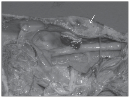
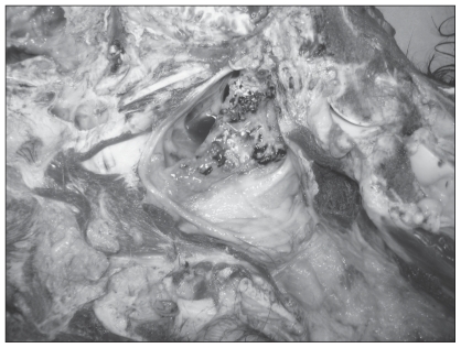
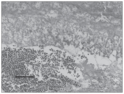
Mycotic encephalitis caused severe ataxia and other neurologic deficits in a horse. The finding of a single, large focus of cerebral malacia, with histopathologic evidence of fungal elements, suggested infection was a result of direct transfer from the frontal sinuses, rather than hematogenous spread from the guttural pouch.
Encéphalite mycotique, ostéomyélite des sinus et mycose de la poche gutturale chez un poulain arabe âgé de 3 ans. Une encéphalite mycotique a causé une ataxie grave et d’autres déficits neurologiques chez un cheval. La découverte d’un grand foyer unique de malacie cérébrale avec une preuve histopathologique d’éléments fongiques a suggéré que l’infection était un transfert direct des sinus frontaux, plutôt qu’une propagation hématogène provenant de la poche gutturale.
(Traduit par Isabelle Vallières)
Figures
Similar articles
-
Progression of mycosis of the auditory tube diverticulum (guttural pouch) after arterial occlusion in a horse with contralateral temporohyoid osteoarthropathy.J Am Vet Med Assoc. 2006 Dec 15;229(12):1945-8. doi: 10.2460/javma.229.12.1945. J Am Vet Med Assoc. 2006. PMID: 17173535
-
Guttural pouch mycosis in a 6-month-old filly.Can Vet J. 2006 Mar;47(3):259-61. Can Vet J. 2006. PMID: 16604984 Free PMC article.
-
A case of guttural pouch mycosis in a horse.Nord Vet Med. 1986 Mar-Apr;38(2):85-9. Nord Vet Med. 1986. PMID: 3725585
-
Update on disorders and treatment of the guttural pouch.Vet Clin North Am Equine Pract. 2015 Apr;31(1):63-89. doi: 10.1016/j.cveq.2014.11.010. Vet Clin North Am Equine Pract. 2015. PMID: 25770066 Review.
-
The management of guttural pouch mycosis.Equine Vet J. 1989 Sep;21(5):321-4. doi: 10.1111/j.2042-3306.1989.tb02679.x. Equine Vet J. 1989. PMID: 2673759 Review. No abstract available.
Cited by
-
Microbiological aspects of osteomyelitis in veterinary medicine: drawing parallels to the infection in human medicine.Vet Q. 2022 Dec;42(1):1-11. doi: 10.1080/01652176.2021.2022244. Vet Q. 2022. PMID: 34936853 Free PMC article. Review.
References
-
- Reed SM, Andrews F. Disorders of the neurologic system. In: Reed SM, Bayly WM, Sellon DC, editors. Equine Internal Medicine. 2nd ed. Philadelphia, Pennsylvania: WB Saunders; 2004. pp. 533–541.
-
- Cook WR. Observations on the aetiology of epistaxis and cranial nerve paralysis in the horse. The Vet Rec. 1966;78:396–406. - PubMed
-
- Lepage OM, Perron MF, Cadore JL. The mystery of fungal infection in the guttural pouches. Vet J. 2004;168:60–64. - PubMed
-
- Ludwig A, Gatineau S, Reynaud MC, et al. Fungal isolation and identication in 21 cases of guttural pouch mycosis in horses (1998–2002) Vet J. 2005;169:457–461. - PubMed
Publication types
MeSH terms
LinkOut - more resources
Full Text Sources
Medical