Retinal thickness after vitrectomy and internal limiting membrane peeling for macular hole and epiretinal membrane
- PMID: 22654493
- PMCID: PMC3363315
- DOI: 10.2147/OPTH.S30288
Retinal thickness after vitrectomy and internal limiting membrane peeling for macular hole and epiretinal membrane
Abstract
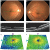
Purpose: To determine the retinal thickness (RT), after vitrectomy with internal limiting membrane (ILM) peeling, for an idiopathic macular hole (MH) or an epiretinal membrane (ERM). Also, to investigate the effect of a dissociated optic nerve fiber layer (DONFL) appearance on RT.
Methods: A non-randomized, retrospective chart review was performed for 159 patients who had successful closure of a MH, with (n = 148), or without (n = 11), ILM peeling. Also studied were 117 patients who had successful removal of an ERM, with (n = 104), or without (n = 13), ILM peeling. The RT of the nine Early Treatment Diabetic Retinopathy Study areas was measured by spectral domain optical coherence tomography (SD-OCT). In the MH-with-ILM peeling and ERM-with-ILM peeling groups, the RT of the operated eyes was compared to the corresponding areas of normal fellow eyes. The inner temporal/inner nasal ratio (TNR) was used to assess the effect of ILM peeling on RT. The effects of DONFL appearance on RT were evaluated in only the MH-with-ILM peeling group.
Results: In the MH-with-ILM peeling group, the central, inner nasal, and outer nasal areas of the retina of operated eyes were significantly thicker than the corresponding areas of normal fellow eyes. In addition, the inner temporal, outer temporal, and inner superior retina was significantly thinner than in the corresponding areas of normal fellow eyes. Similar findings were observed regardless of the presence of a DONFL appearance. In the ERM-with-ILM peeling group, the retina of operated eyes was significantly thicker in all areas, except the inner and outer temporal areas. In the MH-with-ILM peeling group, the TNR was 0.86 in operated eyes, and 0.96 in fellow eyes (P < 0.001). In the ERM-with-ILM peeling group, the TNR was 0.84 in operated eyes, and 0.95 in fellow eyes (P < 0.001). TNR in operated eyes of the MH-without-ILM peeling group was 0.98, which was significantly greater than that of the MH-with-ILM peeling group (P < 0.001). TNR in the operated eyes of the ERM-without-ILM peeling group was 0.98, which was significantly greater than that of ERM-with-ILM peeling group (P < 0.001).
Conclusion: The thinning of the temporal retina and thickening of the nasal retina after ILM peeling does not appear to be disease-specific. In addition, changes in RT after ILM peeling are not related to the presence of a DONFL appearance.
Keywords: epiretinal membrane; internal limiting membrane; macular hole; optical coherence tomography; retinal thickness.
Figures


References
-
- Eckardt C, Eckardt U, Groos S, Luciano L, Reale E. Removal of the internal limiting membrane in macular holes. Clinical and morphological findings. Ophthalmologe. 1997;94:545–551. - PubMed
-
- Olsen TW, Sternberg P, Jr, Capone A, Jr, et al. Macular hole surgery using thrombin-activated fibrinogen and selective removal of the internal limiting membrane. Retina. 1998;18:322–329. - PubMed
-
- Brooks HL., Jr Macular hole surgery with and without internal limiting membrane peeling. Ophthalmology. 2000;107:1939–1948. - PubMed
-
- Kumagai K, Furukawa M, Ogino N, Uemura A, Demizu S, Larson E. Vitreous surgery with and without internal limiting membrane peeling for macular hole repair. Retina. 2004;24:721–727. - PubMed
-
- Christensen UC. Value of internal limiting membrane peeling in surgery for idiopathic macular hole and the correlation between function and retinal morphology. Acta Ophthalmol. 2009;87(Thesis 2):1–23. - PubMed
LinkOut - more resources
Full Text Sources

