Review
. 2012:2012:267120.
doi: 10.1100/2012/267120. Epub 2012 Apr 30.

Distribution of extrasynaptic NMDA receptors on neurons

Affiliations
Review

Distribution of extrasynaptic NMDA receptors on neurons

Ronald S Petralia. ScientificWorldJournal. 2012.

Abstract

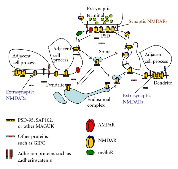
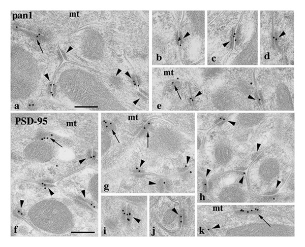
NMDA receptors are found in both synaptic and extrasynaptic locations on neurons. NMDA receptors also can be found on neurons in early stages prior to synaptogenesis, where they may be involved in migration and differentiation. Extrasynaptic NMDA receptors typically are associated with contacts with adjacent processes such as axons and glia. Extrasynaptic NMDA receptor clusters vary in size and may form associations with scaffolding proteins such as PSD-95 and SAP102. The best-characterized extrasynaptic NMDA receptors contain NR1 and NR2B subunits. Extrasynaptic NMDA receptors may be activated by glutamate spillover from synapses or from ectopic release of glutamate. Consequently, extrasynaptic NMDA receptor activation may occur under different circumstances than that for synaptic NMDA receptors, indicating different functional consequences for the neuron. In some cases, activation of extrasynaptic NMDA receptors may have a negative influence on the neuron, leading to cell damage and death, as may occur in some major diseases of the nervous system.


Figures

Figure 1
Figure 1
Immunogold localization of NR1 at the surface (5 nm gold particles indicated by arrows) of axonal growth cone (A; gc) and subsequent presynaptic terminal contact (B; pre) onto dendrites (de) in the CA1 stratum radiatum (A) or stratum oriens (B) of the postnatal day 2 (P2) hippocampus. Growth cones show characteristic collections of large and small endosomal vesicles. A is an axonal growth cone with a few obscure synaptic vesicles near the presynaptic contact, whereas B shows a better developed synapse, including a presynaptic terminal with numerous, distinct synaptic vesicles and high gold labeling on the postsynaptic membrane (asterisk: also, a clathrin-coated pit/vesicle is evident in the dendrite on the other side of the asterisk). The left part of the presynaptic terminal is expanded into a growth cone structure. Scale bars are 100 nm. Reprinted from part of Figure 2 from Wang et al. [14].
Figure 2
Figure 2
Double immunogold labeling of clathrin-coated pits/vesicles (CCP/V; arrowheads) associated with a “bare density” (A; arrow) and extrasynaptic membrane regions (B, C) in the CA1 stratum radiatum of the P2 hippocampus, labeled for NR1 (A, B; 5 nm gold) or NR2A/B (C; 5 nm gold), and clathrin (10 nm gold). The “bare density” (labeled for NR1) on the dendrite in A actually has a fairly close association with an adjacent process. In the dendrite in B, NR1 and clathrin label an early, flat CCP/V adjacent to a CCP/V that is pinching off. In C, NR2A/B labeling on the cell surface is continuous with a clathrin-labeled CCP/V. Scale bar is 100 nm. Reprinted from part of Figure 3 from Petralia et al. [15].
Figure 3
Figure 3
Extrasynaptic NMDARs (NR1 antibody; arrowheads) in the CA1 stratum radiatum of the adult hippocampus (A–H) using immunogold (AB,G,H) or EM immunoperoxidase/DAB (C–F: E and F were processed further with silver/gold toning), and immunofluorescence colocalization of NR2B/VGLUT1/PSD-95/93 (I) or NR2A/VGLUT1/SAP102 (J) in cultured hippocampal neurons. For A–H, extrasynaptic NMDARs can be seen on postsynaptic spines (A–F, H) or dendrites (G; d) adjacent to other processes, including glia (g) and various neuronal processes (see text for details). a: axon terminal; p: presynaptic terminal; asterisk: postsynaptic density. In I, NR2B labeling (red) forms in a perisynaptic ring (**) around synaptic PSD-95/93 (green) and the terminal (labeled with the presynaptic marker, VGLUT1 [blue]) and forms a ring of three puncta around an extrasynaptic punctum of PSD-95/93 (*). In J, this is an enlarged region found along a thin, distal dendrite. Note how NR2A labeling (red) is spread in the perisynaptic regions surrounding two synapses (*): SAP102 (green) forms around the enlargement in conjunction with both the synaptic and extrasynaptic (**) NR2A; the bottom image is a high contrast version of the top one. Scale bars are 100 nm for A–H and 500 nm for I, J. Reprinted from parts of Figures 2, 5, and 6 from Petralia et al. [8].
Figure 4
Figure 4
Diagram illustrating the synaptic and extrasynaptic distributions of NMDARs and associated scaffolding and adhesion proteins, and especially the associations of extrasynaptic NMDARs with adjacent cell processes (see text for details). Although not discussed in this paper, note that other GluRs (AMPARs, kainate and delta iGluRs, and metabotropic GluRs [mGluRs]) are found at synapses and in extrasynaptic locations. AMPARs are typically the most abundant GluRs at synapses and may also be more common than NMDARs in extrasynaptic locations in some neurons. mGluRs are also widespread; some forms are particularly abundant in the perisynaptic zone. Like NMDARs, these GluRs also show close associations with other proteins that affect their trafficking and localization (not illustrated here). The arrangement of different GluRs such as AMPARs and NMDARs within the synapse has been studied but still is not well defined. And although some AMPARs and NMDARs may traffic together in neurons in early development [18], little is known about the association of these different types of GluRs in extrasynaptic locations. For reviews, see Gereau and Swanson [1], Lu and Roche [34], and MacGillavry et al. [35]. Diagram modified from Figure 9 of Petralia et al. [8].
Figure 5
Figure 5
Localization of NMDAR (a–e) and PSD-95 (f–k) antibody labeling mossy terminal glomeruli in the cerebellar granular layer. The pan1 NMDAR antibody is shown here but this was corroborated using 3 more NR1 and 1 NR2A/B antibodies (see Petralia et al. [36] for details). Labeling is limited mainly to synapses (arrows: between the mossy terminal (mt) and dendrite processes from granule cells) and attachment plaques (arrowheads: mainly between granule cell dendrite processes). Scale bar is 200 nm. Reprinted from parts of Figures 2 and 3 from Petralia et al. [36].

References

    1. Gereau RW, IV, Swanson GT. The Glutamate Receptors. Totowa, NJ, USA: Humana Press; 2008.
    1. Kehoe J, Buldakova S, Acher F, Dent J, Bregestovski P, Bradley J. Aplysia cys-loop glutamate-gated chloride channels reveal convergent evolution of ligand specificity. Journal of Molecular Evolution. 2009;69(2):125–141. - PubMed
    1. Janovjak H, Sandoz G, Isacoff EY. A modern ionotropic glutamate receptor with a K+ selectivity signature sequence. Nature Communications. 2011;2(1, article 231) - PubMed
    1. Ryan TJ, Grant SGN. The origin and evolution of synapses. Nature Reviews Neuroscience. 2009;10(10):701–712. - PubMed
    1. Alié A, Manuël M. The backbone of the post-synaptic density originated in a unicellular ancestor of choanoflagellates and metazoans. BMC Evolutionary Biology. 2010;10(1, article 34) - PMC - PubMed

Publication types

Substances

LinkOut - more resources