. 2012 May 28:3:145.
doi: 10.3389/fphys.2012.00145. eCollection 2012.

Emerging role of neuronal exosomes in the central nervous system

Affiliations

Emerging role of neuronal exosomes in the central nervous system

Mathilde Chivet et al. Front Physiol. .

Abstract

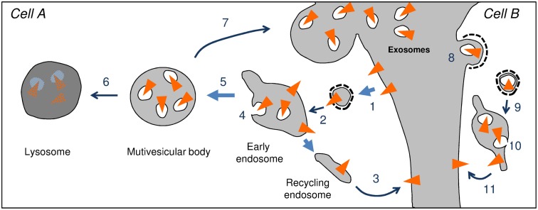
Exosomes are small extracellular vesicles, which stem from endosomes fusing with the plasma membrane, and can be recaptured by receiving cells. They contain lipids, proteins, and RNAs able to modify the physiology of receiving cells. Functioning of the brain relies on intercellular communication between neural cells. These communications can modulate the strength of responses at sparse groups of specific synapses, to modulate circuits underlying associations and memory. Expression of new genes must then follow to stabilize the long-term modifications of the synaptic response. Local changes of the physiology of synapses from one neuron driven by another, have so far been explained by classical signal transduction to modulate transcription, translation, and posttranslational modifications. In vitro evidence now demonstrates that exosomes are released by neurons in a way depending on synaptic activity; these exosomes can be retaken by other neurons suggesting a novel way for inter-neuronal communication. The efficacy of inter-neuronal transfer of biochemical information allowed by exosomes would be far superior to that of direct cell-to-cell contacts or secreted soluble factors. Indeed, lipids, proteins, and RNAs contained in exosomes secreted by emitting neurons could directly modify signal transduction and protein expression in receiving cells. Exosomes could thus represent an ideal mechanism for inter-neuronal transfer of information allowing anterograde and retrograde signaling across synapses necessary for plasticity. They might also allow spreading across the nervous system of pathological proteins like PrPsc, APP fragments, phosphorylated Tau, or Alpha-synuclein.

Keywords: CNS neurons; exosomes; inter-neuronal communication; microvesicles; neurodegeneration; synaptic plasticity.


Figures

Figure 1
Figure 1
Endosomal trafficking of transmembrane proteins (triangle). After endocytosis (1) the endocytic vesicle fuses to early endosomes (2). Proteins can be concentrated into recycling endosomes, which fuse to the plasma membrane and allow re-expression at the cell surface (3). Alternatively proteins can be entrapped in vesicles budding from the limiting membrane of the endosome (4). Maturation of the endosome leads to the individualization of a multivesicular body containing intraluminal vesicles (ILV) (5). The multivesicular body can fuse with lysosomes in which the ILVs and their cargoes are hydrolyzed (6). The multivesicular body can also fuse with the plasma membrane (7) thereby releasing ILVs. Once in the extracellular milieu ILVs are referred to as exosomes. Exosomes released by cell A, can bind to and be endocytosed by a receiving cell [cell B, 8]. The endocytic vesicle containing the exosome fuses with the early endosomes (9). Once inside the endosome, the exosome undergoes back-fusion with the endosomal membrane (10). Fusion of recycling endosomes to the plasma membrane allows expression of protein of the cell A at the surface of cell B. Back-fusion also allows the release of the intraluminal content of exosomes [proteins and RNAs of cell A] into the cytosol of cell B. It is important to note that steps 9, 10, and 11 remain speculative.
Figure 2
Figure 2
(A) Electron micrograph of a multivesicular body present in a neuron of the CA1 region of the adult rat hippocampus. Note the budding of a vesicle from the limiting membrane of the MVB (upper right; Fiona Hemming, unpublished). (B) Electron micrograph of a multivesicular body in a dendrite (colored; CA1 region of an adult rat hippocampus). The protrusion of the dendrite, called dendritic spine, corresponds to the post-synaptic part of a glutamatergic synapse. Two post-synaptic densities, which anchor ionotropic glutamate receptors, are visible. In this case the multivesicular body is present within the dendritic shaft at the base of the spine neck (Fiona Hemming, unpublished).

References

    1. Aguzzi A., Rajendran L. (2009). The transcellular spread of cytosolic amyloids, prions, and prionoids. Neuron 64, 783–79010.1016/j.neuron.2009.12.016 - DOI - PubMed
    1. Arantes R. M., Andrews N. W. (2006). A role for synaptotagmin VII-regulated exocytosis of lysosomes in neurite outgrowth from primary sympathetic neurons. J. Neurosci. 26, 4630–463710.1523/JNEUROSCI.0009-06.2006 - DOI - PMC - PubMed
    1. Babst M. (2011). MVB vesicle formation: ESCRT-dependent, ESCRT-independent and everything in between. Curr. Opin. Cell Biol. 23, 452–45710.1016/j.ceb.2011.04.008 - DOI - PMC - PubMed
    1. Belting M., Wittrup A. (2008). Nanotubes, exosomes, and nucleic acid-binding peptides provide novel mechanisms of intercellular communication in eukaryotic cells: implications in health and disease. J. Cell Biol. 183, 1187–119110.1083/jcb.200810038 - DOI - PMC - PubMed
    1. Brown T. C., Correia S. S., Petrok C. N., Esteban J. A. (2007). Functional compartmentalization of endosomal trafficking for the synaptic delivery of AMPA receptors during long-term potentiation. J. Neurosci. 27, 13311–1331510.1523/JNEUROSCI.2799-07.2007 - DOI - PMC - PubMed

LinkOut - more resources