. 2011 Nov 3:2:60.
doi: 10.3389/fendo.2011.00060. eCollection 2011.

Hypothalamic obesity after craniopharyngioma: mechanisms, diagnosis, and treatment

Affiliations

Hypothalamic obesity after craniopharyngioma: mechanisms, diagnosis, and treatment

Robert H Lustig. Front Endocrinol (Lausanne). .

Abstract

Obesity is a common complication after craniopharyngioma therapy, occurring in up to 75% of survivors. Its weight gain is unlike that of normal obesity, in that it occurs even with caloric restriction, and attempts at lifestyle modification are useless to prevent or treat the obesity. The pathogenesis of this condition involves the inability to transduce afferent hormonal signals of adiposity, in effect mimicking a state of CNS starvation. Efferent sympathetic activity drops, resulting in malaise and reduced energy expenditure, and vagal activity increases, resulting in increased insulin secretion and adipogenesis. Lifestyle intervention is essentially useless in this syndrome, termed "hypothalamic obesity." Pharmacologic treatment is also difficult, consisting of adrenergics to mimic sympathetic activity, or suppression of insulin secretion with octreotide, or both. Recently, bariatric surgery (Roux-en-Y gastric bypass, laparoscopic gastric banding, truncal vagotomy) have also been attempted with variable results. Early and intensive management is required to mitigate the obesity and its negative consequences.

Keywords: craniopharyngioma; ghrelin; hypothalamic obesity; insulin; leptin resistance; octreotide; symapthetic nervous system; vagus nerve.


Figures

Figure 1
Figure 1
Neuroendocrine regulation of energy balance. The afferent system: neural (e.g., vagal) and hormonal (ghrelin, insulin, leptin) signals are generated from the liver, gut, pancreas, and adipose. In addition, norepinephrine from the locus cœruleus and serotonin (5-HT) from the median raphe are elaborated. These signals of satiety vs. hunger, and thinness vs. fatness are interpreted in the ventromedial hypothalamus (VMH). These signals are then integrated in the paraventricular nucleus (PVN) and lateral hypothalamus (LHA). The efferent system: efferent signals from these areas in turn stimulate the sympathetic nervous system (SNS) to expend energy by activating β3-adrenergic receptors and uncoupling proteins in the adipocyte, to release energy the form of lipolysis, heat, or physical activity. Conversely, the parasympathetic nervous system (efferent vagal) increases insulin secretion, with resultant adipogenesis and energy storage, and also increases insulin sensitivity through direct effects on the adipose tissue (Lustig, 2006). From Nature Publishing Group, with permission.
Figure 2
Figure 2
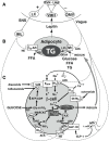
Central regulation of leptin signaling, autonomic innervation of the adipocyte and β-cell, and the starvation response. (A) The arcuate nucleus transduces the peripheral leptin signal as one of sufficiency or deficiency. In leptin sufficiency, efferents from the hypothalamus synapse in the locus coeruleus, which stimulates the sympathetic nervous system. In leptin deficiency, efferents from the hypothalamus stimulate the dorsal motor nucleus of the vagus. (B) Autonomic innervation and hormonal stimulation of white adipose tissue. In leptin sufficiency, norepinephrine binds to the β3-adrenergic receptor, which stimulates hormone-sensitive lipase, promoting lipolysis of stored triglyceride into free fatty acids. In leptin deficiency, vagal acetylcholine increases adipose tissue insulin sensitivity (documented only in rats to date), promotes uptake of glucose and free fatty acids for lipogenesis, and promotes triglyceride uptake through activation of lipoprotein lipase. (C) Autonomic innervation and hormonal stimulation of the β-cell. Glucose entering the cell is converted to glucose-6-phosphate by the enzyme glucokinase, generating ATP, which closes an ATP-dependent potassium channel, resulting in cell depolarization. A voltage-gated calcium channel opens, allowing for intracellular calcium influx, which activates neurosecretory mechanisms leading to insulin vesicular exocytosis. In leptin sufficiency, norepinephrine binds to α2-adrenoceptors on the β-cell membrane to stimulate inhibitory G proteins, decrease adenyl cyclase and its product cAMP, and thereby reduce protein kinase A levels and insulin release. In leptin deficiency, the vagus stimulates insulin secretion through three mechanisms. First, acetylcholine binds to a M3 muscarinic receptor, opening a sodium channel, which augments the ATP-dependent cell depolarization, increasing the calcium influx, and insulin exocytosis. Secondly, acetylcholine activates a pathway that increases protein kinase C, which also promotes insulin secretion. Thirdly, the vagus innervates L-cells of the small intestine, which secrete glucagon-like peptide-1, which activates protein kinase A, contributing to insulin exocytosis. Octreotide binds to a somatostatin receptor on the β-cell, which is coupled to the voltage-gated calcium channel, limiting calcium influx and the amount of insulin released in response to glucose. (Lustig, ; reprinted with kind permission of Humana, Totowa, NJ, USA). α2-AR, α2-adrenergic receptor; β3-AR, β3-adrenergic receptor; AC, adenyl cyclase; ACh, acetylcholine; DAG, diacylglycerol; DMV, dorsal motor nucleus of the vagus; FFA, free fatty acids; Gi, inhibitory G protein; GK, glucokinase; GLP-1, glucagon-like peptide-1; GLP-1R, GLP-1 receptor; Glu-6-PO4, glucose-6-phosphate; Glut4, glucose transporter-4; HSL, hormone-sensitive lipase; IML, intermediolateral cell column; IP3, inositol triphosphate; LC, locus coeruleus; LHA, lateral hypothalamic area; LPL, lipoprotein lipase; MARCKS, myristoylated alanine-rich protein kinase C substrate; NE, norepinephrine; PIP2, phosphatidylinositol; PKA, protein kinase A; PKC, protein kinase C; PLC, phospholipase C; PVN, paraventricular nucleus; SSTR5, somatostatin-5 receptor; TG, triglyceride; VCa, voltage-gated calcium channel; VMH, ventromedial hypothalamus; SUR, sufonylurea receptor (Lustig, 2006). From Nature Publishing Group, with permission.
Figure 3
Figure 3
Scatterplot of insulin secretion (Corrected Insulin Response, or CIRgp) vs. sensitivity (Composite Insulin Sensitivity Index, or CISI) plotted logarithmically in 113 obese non-diabetic children. A negative linear correlation was noted (r = −0.54, p < 0.001). Different racial and etiopathogenic groups tended to plot in different areas. Arbitrary cutoffs (dashed lines) for CIRgp (1.5) and CISI (1.7) divide the plot into four quadrants. The majority of Caucasian children (open squares) plotted in the lower right quadrant, with a CIRgp less than 1.5 and a CISI greater than 1.7, indicating lower insulin secretion and better insulin sensitivity. The preponderance of children with hypothalamic obesity (gray squares) plotted in the upper right quadrant, with a CIRgp of greater than 1.5, and with a CISI of greater than 1.7, indicating insulin hypersecretion with better insulin sensitivity. Finally the majority of African American children (filled squares) plotted in the upper left quadrant, with a CIRgp of greater than 1.5 and a CISI of less than 1.7, indicating both insulin hypersecretion and resistance (Preeyasombat et al., 2005). From Elsevier, with permission.

References

    1. al-Adsani H., Hoffer L. J., Silva J. E. (1997). Resting energy expenditure is sensitive to small dose changes in patients on chronic thyroid hormone replacement. J. Clin. Endocrinol. Metab. 82, 1118–112510.1210/jc.82.4.1118 - DOI - PubMed
    1. Arbuzova A., Murray D., McLaughlin S. (1998). MARCKS, membranes, and calmodulin: kinetics of their interaction. Biochim. Biophys. Acta 1376, 369–379 - PubMed
    1. Babinski M. J. (1900). Tumeur du corps pituitaire san acromegalie et avec arret de developpement des organes genitaux. Rev. Neurol. 8, 531–533
    1. Balthasar N., Dalgaard L. T., Lee C. E., Yu J., Funahashi H., Williams T., Ferreira M., Tang V., McGovern R. A., Kenny C. D., Christiansen L. M., Edelstein E., Choi B., Boss O., Aschkenasi C., Zhang C. Y., Mountjoy K., Kishi T., Elmquist J. K., Lowell B. B. (2005). Divergence of melanocortin pathways in the control of food intake and energy expenditure. Cell 123, 493–50510.1016/j.cell.2005.08.035 - DOI - PubMed
    1. Berthoud H. R., Jeanrenaud B. (1979). Acute hyperinsulinemia and its reversal by vagotomy following lesions of the ventromedial hypothalamus in anesthetized rats. Endocrinology 105, 146–15110.1210/endo-105-1-146 - DOI - PubMed