. 2011 Nov 29:2:87.
doi: 10.3389/fendo.2011.00087. eCollection 2011.

Differentiation of Human Adipose-Derived Stem Cells into "Brite" (Brown-in-White) Adipocytes

Affiliations

Differentiation of Human Adipose-Derived Stem Cells into "Brite" (Brown-in-White) Adipocytes

Didier F Pisani et al. Front Endocrinol (Lausanne). .

Abstract

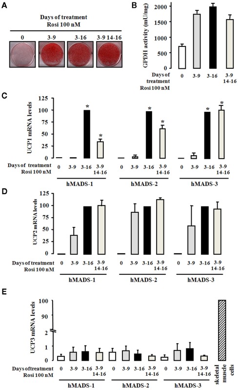
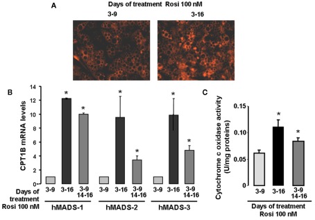
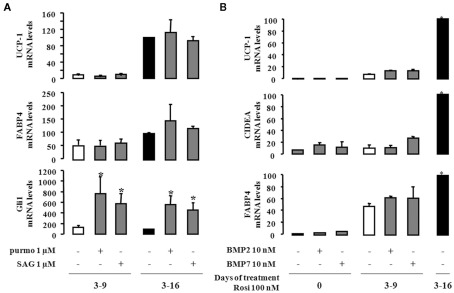
It is well established now that adult humans possess active brown adipose tissue (BAT) which represents a potential pharmacological target to combat obesity and associated diseases. Moreover thermogenic brown-like adipocytes ("brite adipocytes") appear also in mouse white adipose tissue (WAT) upon β3-adrenergic stimulation. We had previously shown that human multipotent adipose-derived stem cells (hMADS) are able to differentiate into cells which exhibit the key properties of human white adipocytes, and then to convert into functional brown adipocytes upon PPARγ activation. In light of a wealth of data indicating that thermogenic adipocytes from BAT and WAT have a distinct cellular origin, we have characterized at the molecular level UCP1 positive hMADS adipocytes from both sexes as brite adipocytes. Conversion of white to brown hMADS adipocytes is dependent on PPARγ activation with rosiglitazone as the most potent agonist and is inhibited by a PPARγ antagonist. In contrast to mouse cellular models, hMADS cells conversion into brown adipocytes is weakly induced by BMP7 treatment and not modulated by activation of the Hedgehog pathway. So far no primary or clonal precursor cells of human brown adipocytes have been obtained that can be used as a tool to develop therapeutic drugs and to gain further insights into the molecular mechanisms of brown adipogenesis in humans. Thus hMADS cells represent a suitable human cell model to delineate the formation and/or the uncoupling capacity of brown/brite adipocytes that could help to dissipate caloric excess intake among individuals.

Keywords: BAT; UCP1; WAT; adipocyte; brite adipocyte; differentiation; rosiglitazone; stem cells.


Figures

Figure 1
Figure 1
Conversion of hMADS cells into brown- like adipocytes. hMADS-1, 2, and 3 cells were induced to differentiate into adipocytes as described under the Section “Materials and Methods.” Hundred nanometer rosiglitazone was added during the indicated periods of treatment. At day 16, (A) cells were fixed and stained by Oil Red O, (B) GPDH activity was determined, and (C–E) UCP1, UCP2, and UCP3 mRNA levels, respectively, were measured by quantitative RT-PCR. Results are mean ± SEM of three (B,C) or two (D,E) independent experiments. Results in (C) and (D) are expressed by taking as 100% the value obtained for rosiglitazone treatment between days 3 and 16. In (E), human skeletal muscle cells were used as positive control for UCP3 expression. Results in (E) are expressed taking as 100% the value obtained for skeletal muscle cells. *p < 0.05 vs. cells treated between days 3 and 9.
Figure 2
Figure 2
Activation of PPARγ is involved in the conversion of white to brown adipocytes. hMADS-2 cells were induced to differentiate into white adipocytes in the presence of 100 nM rosiglitazone between days 3 and 9, and then treated with various component as indicated between days 14 and 16. (A) Effect of various PPARγ agonists (Rosiglitazone, Troglitazone, and Ciglitazone) analyzed by quantitative RT-PCR of UCP1 gene expression. (B) Effect of the PPARγ antagonist GW9662 on UCP1 expression stimulated by rosiglitazone. Results are the mean ± SEM of two independent experiments. Results are expressed by taking as 100% the value obtained at 100 nM rosiglitazone. *p < 0.05 vs. cells exposed to 100 nM rosiglitazone between days 3–9 and days 14–16.
Figure 3
Figure 3
Mitochondriogenesis of hMADS cells after conversion to brown-like adipocytes. hMADS-2 cells were induced to differentiate into adipocytes in the presence of 100 nM rosiglitazone between days 3 and 9 or between days 3 and 16. Alternatively, after removal at day 9, rosiglitazone was re-added or not at day 14 for 2 days. (A) Mitochondria visualized by epifluorescence in living cells in the presence of the mitotracker dye, (B) CPT1B mRNA levels determined at day 16 by quantitative RT-PCR in hMADS-1, 2, and 3 cells and (C) Cytochrome c oxidase activity determined at day 16. Results are mean ± SEM (B,C) and are representative experiments (A) of three independent experiments performed on different series of cells. *p < 0.05 vs. cells treated between days 3 and 9.
Figure 4
Figure 4
Effect of Hedgehog and BMP signaling in the conversion of white to brown adipocytes. hMADS-2 cells were induced to differentiate into adipocytes in presence of 100 nM rosiglitazone at the indicate times. (A), Purmorphamine (purmo) or smoothened agonist (SAG) as effectors of the Hedgehog signaling pathway were added between days 14 and 18. Their effects were analyzed by measuring UCP1, FABP4, and Gli1 mRNA expression. (B) hMADS-2 cells were treated or not with 10 nM BMP2 or BMP7 between days 0 and 16, and analyzed at day 16. Results are the mean ± SEM of three independent experiments and are expressed by taking as 100% the value obtained for 100 nM rosiglitazone treatment between days 14 and 18. *p < 0.05 vs. cells treated only by rosiglitazone 100 nM.

References

    1. Ailhaud G., Grimaldi P., Negrel R. (1992). Cellular and molecular aspects of adipose tissue development. Annu. Rev. Nutr. 12, 207–23310.1146/annurev.nu.12.070192.001231 - DOI - PubMed
    1. Barbatelli G., Murano I., Madsen L., Hao Q., Jimenez M., Kristiansen K., Giacobino J. P., De Matteis R., Cinti S. (2010). The emergence of cold-induced brown adipocytes in mouse white fat depots is determined predominantly by white to brown adipocyte transdifferentiation. Am. J. Physiol. Endocrinol. Metab. 298, E1244–E125310.1152/ajpendo.00600.2009 - DOI - PubMed
    1. Bezy O., Elabd C., Cochet O., Petersen R. K., Kristiansen K., Dani C., Ailhaud G., Amri E. Z. (2005). Delta-interacting protein A, a new inhibitory partner of CCAAT/enhancer-binding protein beta, implicated in adipocyte differentiation. J. Biol. Chem. 280, 11432–1143810.1074/jbc.M411741200 - DOI - PubMed
    1. Cinti S. (2009a). Reversible physiological transdifferentiation in the adipose organ. Proc. Nutr. Soc. 68, 340–34910.1017/S0029665109990140 - DOI - PubMed
    1. Cinti S. (2009b). Transdifferentiation properties of adipocytes in the adipose organ. Am. J. Physiol. Endocrinol. Metab. 297, E977–E98610.1152/ajpendo.00183.2009 - DOI - PubMed

LinkOut - more resources